


 全部商品分类
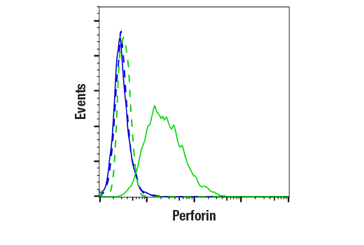


 用小程序,查商品更便捷


Monoclonal antibody is produced by immunizing animals with recombinant protein specific to mouse perforin protein.

Product Usage Information
| Application | Dilution |
|---|---|
| Western Blotting | 1:1000 |
| Immunofluorescence (Frozen) | 1:50 - 1:100 |
| Immunofluorescence (Immunocytochemistry) | 1:50 - 1:100 |
| Flow Cytometry (Fixed/Permeabilized) | 1:50 - 1:200 |
Specificity/Sensitivity
Species Reactivity:
Mouse
Supplied in 10 mM sodium HEPES (pH 7.5), 150 mM NaCl, 100 µg/ml BSA, 50% glycerol and less than 0.02% sodium azide. Store at –20°C. Do not aliquot the antibody.
参考图片
Flow cytometric analysis of A20 cells (blue, negative) and CTLL-2 cells (green, positive) using Perforin (E9F7N) Rabbit mAb (solid lines) or a concentration-matched Rabbit (DA1E) mAb IgG XP® Isotype Control #3900 (dashed lines). Anti-rabbit IgG (H+L), F(ab')2 Fragment (Alexa Fluor® 488 Conjugate) #4412 was used as a secondary antibody.
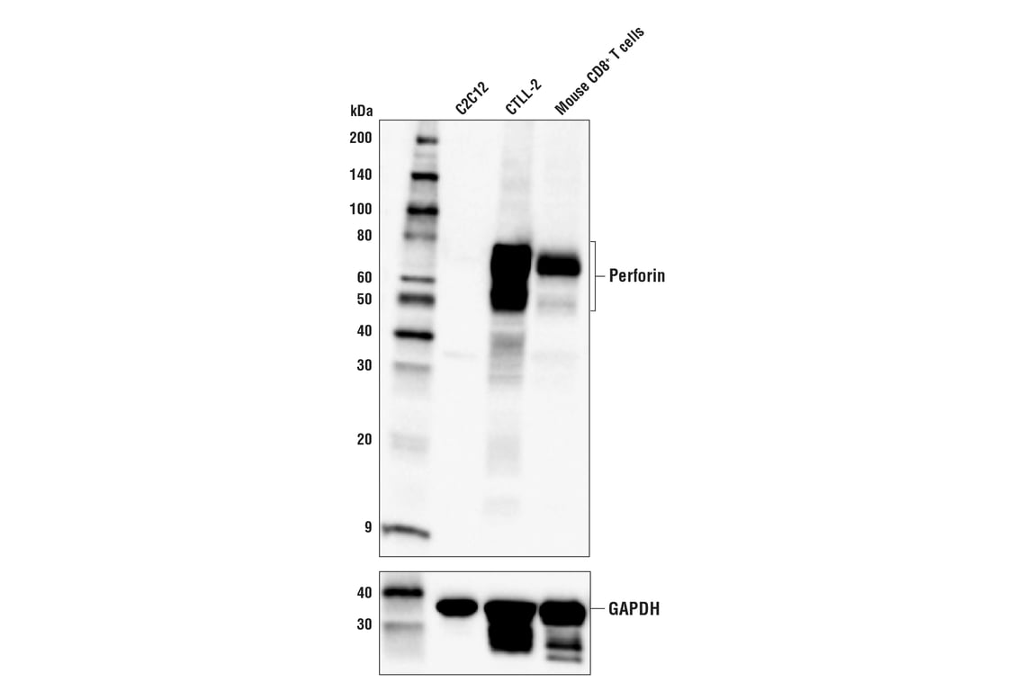
Western blot analysis of extracts from C2C12 cells, CTLL-2 cells, and mouse activated CD8+ T cells using Perforin (E9F7N) Rabbit mAb (upper) or GAPDH (D16H11) XP® Rabbit mAb #5174 (lower).
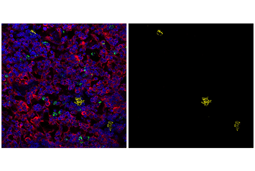
Confocal immunofluorescent analysis of mouse spleen using Perforin (E9F7N) Rabbit mAb (yellow), CD4 (RPA-T4) Mouse mAb (FITC Conjugate) #48705 (green), and S6 Ribosomal Protein (54D2) Mouse mAb (Alexa Fluor® 647 Conjugate) #5548 (red). Samples were mounted in ProLong® Gold Antifade Reagent with DAPI #8961 (blue).
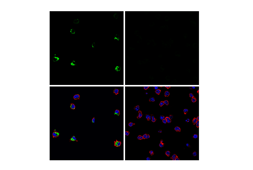
Confocal immunofluorescent analysis of CTLL-2 cells (left, positive) or BA/F3 cells (right, negative) using Perforin (E9F7N) Rabbit mAb (green). Actin filaments were labeled with DyLight™ 554 Phalloidin #13054 (red). Samples were mounted in ProLong® Gold Antifade Reagent with DAPI #8961 (blue).