


 全部商品分类


1/2

品牌: BD Pharmingen
 下载产品说明书 下载SDS
 用小程序,查商品更便捷


 收藏
收藏
 对比
对比 咨询
咨询反应种属:
Human (QC Testing), Cow (Reported)
来源宿主:
Mouse BALB/c IgG2b, κ
产品介绍
产品介绍
产品信息
荧光素标记
抗原名称
Perforin

宿主
Mouse BALB/c IgG2b, κ
免疫原
Purified Granules from the Human Lymphoma Cell Line YT
简单描述
Perforin has a key role in cell-mediated cytotoxicity. It is a 70 kDa cytolytic protein that is expressed in the cytoplasmic granules of cytotoxic T lymphocytes (CTLs) and natural killer (NK) cells. CTLs are involved in eliminating virally infected cells, in anti-tumor immune responses, in allograft rejections, and in some autoimmune diseases. NK cells are important for tumor surveillance and destruction and are involved in allograft rejections. Cytotoxic cells release the contents of their cytotoxic granules, including perforin upon recognition of their target cell. In the presence of calcium, perforin forms transmembrane channels or pores in the membrane of the target cell leading to a cell death that resembles apoptosis. The ability to detect perforin-positive cells with specific antibody should be useful in identifying and understanding perforin-mediated reactions.
Clone δG9 reacts with human and bovine perforin. It does not cross-react with mouse perforin. Purified granules from the human lymphoma cell line YT were used as immunogen. Clone δG9 was initially characterized by immunoprecipitation and immunohistochemistry of frozen tissue sections. The antibody stains scattered lymphocytes in red pulp of spleen, and scattered infiltrated lymphocytes in lymphoma.
商品描述
δG9
Perforin has a key role in cell-mediated cytotoxicity. It is a 70 kDa cytolytic protein that is expressed in the cytoplasmic granules of cytotoxic T lymphocytes (CTLs) and natural killer (NK) cells. CTLs are involved in eliminating virally infected cells, in anti-tumor immune responses, in allograft rejections, and in some autoimmune diseases. NK cells are important for tumor surveillance and destruction and are involved in allograft rejections. Cytotoxic cells release the contents of their cytotoxic granules, including perforin upon recognition of their target cell. In the presence of calcium, perforin forms transmembrane channels or pores in the membrane of the target cell leading to a cell death that resembles apoptosis. The ability to detect perforin-positive cells with specific antibody should be useful in identifying and understanding perforin-mediated reactions.
Clone δG9 reacts with human and bovine perforin. It does not cross-react with mouse perforin. Purified granules from the human lymphoma cell line YT were used as immunogen. Clone δG9 was initially characterized by immunoprecipitation and immunohistochemistry of frozen tissue sections. The antibody stains scattered lymphocytes in red pulp of spleen, and scattered infiltrated lymphocytes in lymphoma.
同种型
Mouse BALB/c IgG2b, κ
克隆号
克隆 δG9 (RUO)
产品详情
PerCP-Cy5.5
PerCP-Cy5.5 dye is part of the BD blue family of dyes. This tandem fluorochrome is comprised of a fluorescent protein complex (PerCP) with an excitation maximum (Ex Max) of 482 nm and an acceptor dye with an emission maximum (Em Max) at 676 nm. PerCP-Cy5 is designed to be excited by the blue laser (488-nm) and detected using an optical filter centered near 680 nm (e.g., a 695/40 nm bandpass filter). The donor dye can be partially excited by the Violet (405-nm) laser resulting in cross-laser excitation and fluorescence spillover. Please ensure that your instrument’s configurations (lasers and optical filters) are appropriate for this dye.

PerCP-Cy5.5
Blue 488 nm
482 nm
676 nm
应用
实验应用
Intracellular staining (flow cytometry) (Routinely Tested)
推荐用量
5 µl
反应种属
Human (QC Testing), Cow (Reported)
目标/特异性
Perforin
背景
别名
PRF1; P1; PERF; PFN1; PFP; Perforin-1; Cytolysin; FLH2; HPLH2
制备和贮存
存储溶液
Aqueous buffered solution containing BSA and ≤0.09% sodium azide.
保存方式
Aqueous buffered solution containing BSA and ≤0.09% sodium azide.
文献
文献
研发参考(7)
1. Endsley JJ, Furrer JL, Endsley MA, et al. Characterization of bovine homologues of granulysin and NK-lysin. J Immunol. 2004; 173(4):2607-2614. (Clone-specific: Flow cytometry, Western blot).
2. Fox WM 3rd, Hameed A, Hutchins GM, et al. Perforin expression localizing cytotoxic lymphocytes in the intimas of coronary arteries with transplant-related accelerated arteriosclerosis. Hum Pathol. 1993; 24(5):477-482. (Clone-specific: Immunohistochemistry).
3. Hameed A, Fox WM, Kurman RJ, Hruban RH, Podack ER. Perforin expression in endometrium during the menstrual cycle. Int J Gynecol Pathol. 1995; 14(2):143-150. (Clone-specific: Flow cytometry).
4. Hameed A, Fox WM, Kurman RJ, Hruban RH, Podack ER. Perforin expression in human cell-mediated luteolysis. Int J Gynecol Pathol. 1995; 14(2):151-157. (Clone-specific: Immunohistochemistry).
5. Hameed A, Olsen KJ, Cheng L, Fox WM 3rd, Hruban RH, Podack ER. Immunohistochemical identification of cytotoxic lymphocytes using human perforin monoclonal antibody. Am J Pathol. 1992; 140(5):1025-1030. (Immunogen: Immunohistochemistry, Immunoprecipitation).
6. Hameed A, Podack ER, Fox WM, Schafer RW, Sherman ME. Detection of perforin in human peritoneal fluid T-lymphocytes. Acta Cytol. 1996; 40(3):401-407. (Clone-specific: Immunohistochemistry).
7. Rukavina D, Balen-Marunic S, Rubesa G, Orlic P, Vujaklija K, Podack ER. Perforin expression in peripheral blood lymphocytes in rejecting and tolerant kidney transplant recipients. Transplantation. 1996; 61(2):285-291. (Clone-specific: Flow cytometry).
参考图片
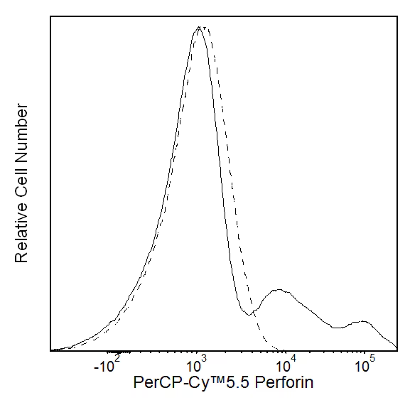
Flow cytometric analysis of perforin expression in human peripheral blood mononuclear cells. Human peripheral blood mononuclear cells were fixed and permeabilized with BD Cytofix/Cytoperm™ Fixation and Permeabilization Solution (Cat. No. 554722). The cells were then washed with and stained in BD Perm/Wash™ Buffer (Cat. No. 554723) with either PerCP-Cy™5.5 Mouse IgG2b, κ Isotype Control (Cat. No. 558304; dashed line histogram) or PerCP-Cy™5.5 Mouse Anti-Human Perforin antibody (Cat. No. 563762; solid line histogram). The fluorescence histograms were derived from gated events with the forward and side light-scatter characteristics of intact lymphocytes. Flow cytometric analysis was performed using a BD™ LSR II Flow Cytometer System.
声明 :本官网所有报价均为常温或者蓝冰运输价格,如有产品需要干冰运输,需另外加收干冰运输费。