BD Phosflow™ Perm Buffer IV (Perm Buffer IV) is intended for the permeabilization of paraformaldehyde-fixed single-cell suspensions prepared from lymphoid tissues, peripheral blood mononuclear cells (PBMC) and lysed whole blood cells. Permeabilization of cells by treatment with this buffer and subsequent washing with staining buffer enables access for fluorescent antibodies to stain intracellular molecules. This buffer preserves the light scattering properties of cells while providing the capacity for intracellular immunofluorescent staining in preparation for Phosflow cytometric analysis.
Perm Buffer IV is formulated for improved immunofluorescent staining and detection of certain intracellular phosphorylated proteins. PBMC, for example, can be treated with protein kinase activators (eg, phorbol esters) or inhibitors, mitogens or cytokines and fixed with BD
Cytofix™ Fixation Buffer (Cat. No. 554655). In the case of peripheral whole blood cells, the blood cells can be treated, erythrocytes lysed and the remaining leukocytes fixed using BD Phosflow™ Lyse/Fix Buffer (Cat. No. 558049). Following fixation, the cells can then be permeabilized with Perm Buffer IV and washed with staining buffer to allow for effective intracellular staining. Designed for optimal staining of cells destined for flow cytometric analysis, Perm Buffer IV can be used with fluorescent antibodies specific for phosphorylated cell signaling molecules to define the nature of signaling pathways induced by individual cells responding to various activators, inhibitors, or combinations thereof. At a 1.0× concentration, Perm Buffer IV has been shown to support large fold changes (i.e., mean fluorescent intensity staining ratios of treated versus untreated cell populations) for phosphorylated Signal transducer and activators of transcription (Stat) proteins. However, it should be noted that at a 1.0× concentration, Perm Buffer IV may result in a greater cell loss (~18% to 48%) than other BD permeabilization buffers and decreased ability to stain certain cell-surface markers. At a 0.5× concentration, the BD Phosflow™ Perm Buffer IV has been shown to be compatible with the immunofluorescent staining of phosphorylated intracellular Stat proteins, although sometimes with lower fold changes. Unlike the 1.0× concentration of Perm Buffer IV, the 0.5× concentration of Buffer IV offers improved compatibility for staining cell-surface CD markers and results in comparable cell recoveries when compared with the BD Phosflow™ Perm Buffers I, II, and III. The figures demonstrate some examples of intracellular and cell-surface staining of cells that were permeabilized with Perm Buffer IV at 1× and 0.5× concentrations.
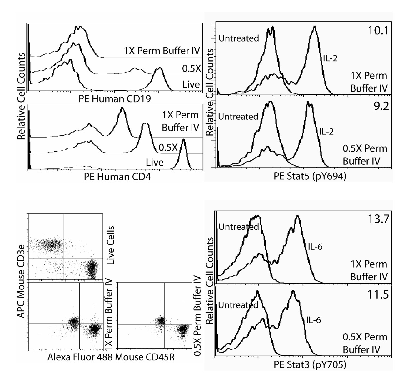
Immunofluorescent staining of human and mouse lymphocytes after permeabilization with Perm Buffer IV. Human whole blood (top row) and mouse splenocytes (bottom row) were treated with 100 ng/ml IL-2 (Cat. No. 554603, top right figure) or 100 ng/ml IL-6 (Cat. No. 550071, bottom right figure) or left untreated (all 4 figures) for 15 minutes. Erythrocytes were lysed and leukocytes were fixed and permeabilized with Perm Buffer IV at either 1× or 0.5× according to the Recommended Assay Procedure. Then the human cells were stained with PE Mouse Anti-Human CD19 (Cat. No. 555413, top panel of top left figure), PE Mouse Anti-Human CD4 (Cat. No. 347327, bottom panel of top left figure), or PE Mouse Anti-Stat5 (pY694) (Cat. No. 612567, top right figure). Similarly, the mouse cells were stained with APC Hamster Anti-Mouse CD3e (Cat. No. 553066) and Alexa Fluor® 488 Rat Anti-Mouse CD45R (Cat. No. 557669) in the presence of Purified Rat Anti-Mouse CD16/CD32 (Mouse BD Fc Block™, Cat. No. 553142, bottom left figure) or PE Mouse Anti-Stat3 (pY705) (Cat. No. 612569, bottom right figure). Additional controls for the cell-surface staining were live cells that had undergone erythrocyte lysis using BD Pharm Lyse™ lysing buffer (Cat. No. 555899, left column) but no fixation or permeabilization. The fold change is indicated for staining of Stat5 (pY694) or Stat3 (pY705) with each perm buffer concentration (upper right corners of panels in the right column). Lymphocytes were selected by scatter profile. Flow cytometry was performed on a BD FACSCanto™ II flow cytometer.


 全部商品分类


 下载产品说明书
 用小程序,查商品更便捷


 收藏
收藏
 对比
对比 咨询
咨询