


 全部商品分类


BD Cytoperm™ Permeabilization Buffer Plus

 下载产品说明书
 用小程序,查商品更便捷


 收藏
收藏
 对比
对比 咨询
咨询
参考图片
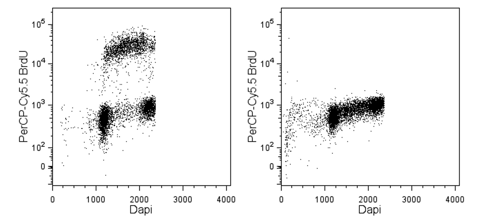
Flow cytometric analysis of DNA synthesis by TK-1 cells. TK-1 cells were either pulsed with 50 µM BrdU for 1 hour (left panel) or were not pulsed (right panel). Staining was performed using BD Cytoperm™ Permeabilization Buffer Plus in the procedure from the BD Pharmingen™ FITC and APC BrdU Flow Kits. The permeabilized cells were stained with the PerCP-Cy™5.5 Mouse Anti-BrdU monoclonal antibody (Cat. No. 560809) followed by the DNA-specific dye, DAPI dihydrochloride at 1 µg/mL (Sigma, Cat. No. D9542). Two-color flow cytometric dot plots showing the correlated expression patterns of DAPI vs BrdU were derived from gated events with the forward and side light-scatter characteristics of viable lymphocytes. Flow cytometry was performed with doublet discrimination using a BD™ LSRII system.
Flow cytometric analysis of DNA synthesis by TK-1 cells. TK-1 cells were either pulsed with 50 µM BrdU for 1 hour (left panel) or were not pulsed (right panel). Staining was performed using BD Cytoperm™ Permeabilization Buffer Plus in the procedure from the BD Pharmingen™ FITC and APC BrdU Flow Kits. The permeabilized cells were stained with the PerCP-Cy™5.5 Mouse Anti-BrdU monoclonal antibody (Cat. No. 560809) followed by the DNA-specific dye, DAPI dihydrochloride at 1 µg/mL (Sigma, Cat. No. D9542). Two-color flow cytometric dot plots showing the correlated expression patterns of DAPI vs BrdU were derived from gated events with the forward and side light-scatter characteristics of viable lymphocytes. Flow cytometry was performed with doublet discrimination using a BD™ LSRII system.