


 全部商品分类


Phospho-Akt (Ser473) Antibody

 下载产品说明书 下载COA 下载SDS
 用小程序,查商品更便捷


 收藏
收藏
 对比
对比 咨询
咨询
Polyclonal antibodies are produced by immunizing animals with a synthetic phosphopeptide corresponding to residues surrounding Ser473 of mouse Akt. Antibodies are purified by protein A and peptide affinity chromatography.
Product Usage Information
| Application | Dilution |
|---|---|
| Western Blotting | 1:1000 |
| Immunoprecipitation | 1:100 |
| Immunofluorescence (Immunocytochemistry) | 1:50 - 1:200 |
| Flow Cytometry (Fixed/Permeabilized) | 1:100 - 1:400 |
Specificity/Sensitivity
Species Reactivity:
Human, Mouse, Rat, Hamster, Monkey, D. melanogaster, Bovine, Dog
Supplied in 10 mM sodium HEPES (pH 7.5), 150 mM NaCl, 100 µg/ml BSA and 50% glycerol. Store at –20°C. Do not aliquot the antibody.
参考图片
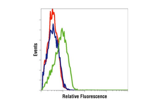
Flow cytometric analysis of LNCaP cells, untreated (green) or LY294002-treated (blue), using Phospho-Akt (Ser473) Antibody compared to a nonspecific negative control antibody (red).
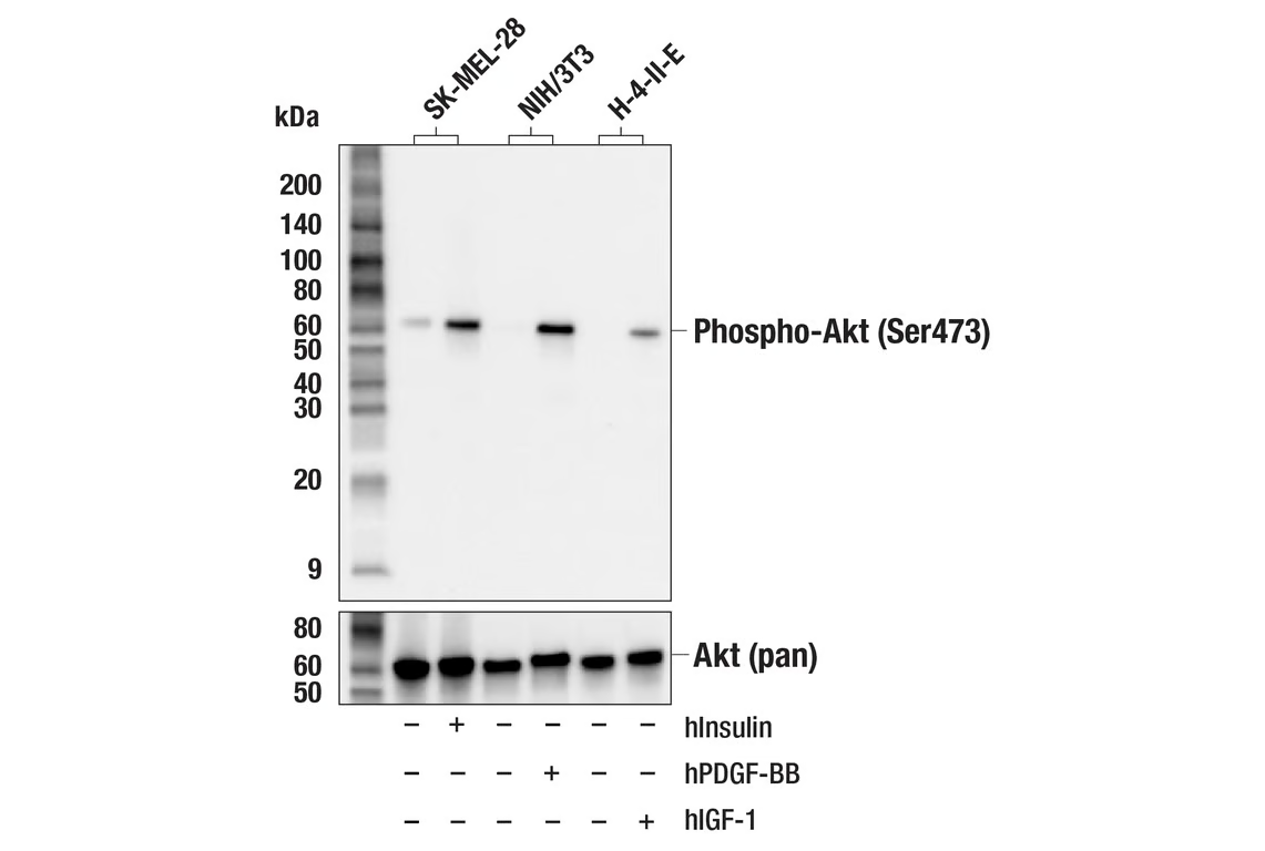
Western blot analysis of extracts from SK-MEL-28, NIH/3T3, and H-4-II-E cells, untreated (-) or treated (+) as indicated with: human Insulin (hInsulin; 100 nM, 20 min), mouse PDGF-BB (mPDGF-BB; 100 ng/mL, 20 min), or human IGF-1 (hIGF-1; 100 ng/mL, 5 min) using Phospho-Akt (Ser473) Antibody (upper) or Akt (pan) (C67E7) Rabbit mAb #4691 (lower).
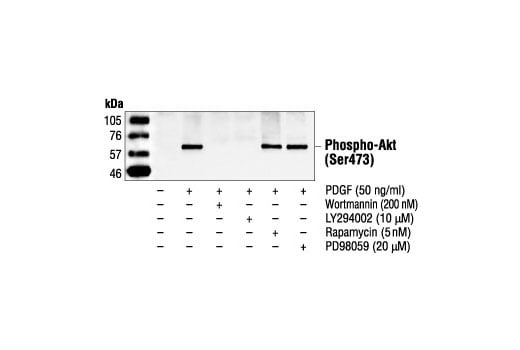
Western blot analysis of extracts from NIH/3T3 cells, untreated or treated with PDGF, wortmannin, LY294002, rapamycin or PD98059, using Phospho-Akt (Ser473) Antibody.
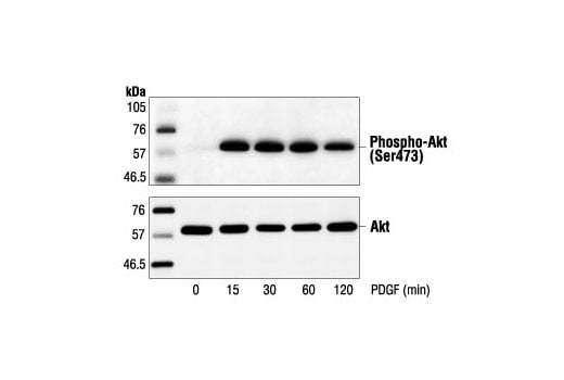
Western blot analysis of extracts from NIH/3T3 cells, untreated or treated with PDGF for the indicated times, using Phospho-Akt (Ser473) Antibody (upper) or Akt Antibody #9272 (lower).
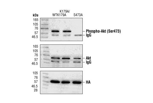
Western blot analysis of immunoprecipitated Akt from 293 cells transiently transfected with HA-tagged Akt (WT), HA-tagged K179A mutant Akt and HA-tagged K179A/S473A mutant Akt, using Phospho-Akt (Ser473) Antibody (upper), Akt antibody (middle) or HA antibody (lower). Phospho-Akt (Ser473) Antibody does not recognize Akt with an alanine substituion at Ser473. (Polakiewicz, R.D. et al. [1998] J. Biol. Chem. 273, 23534-23541.)
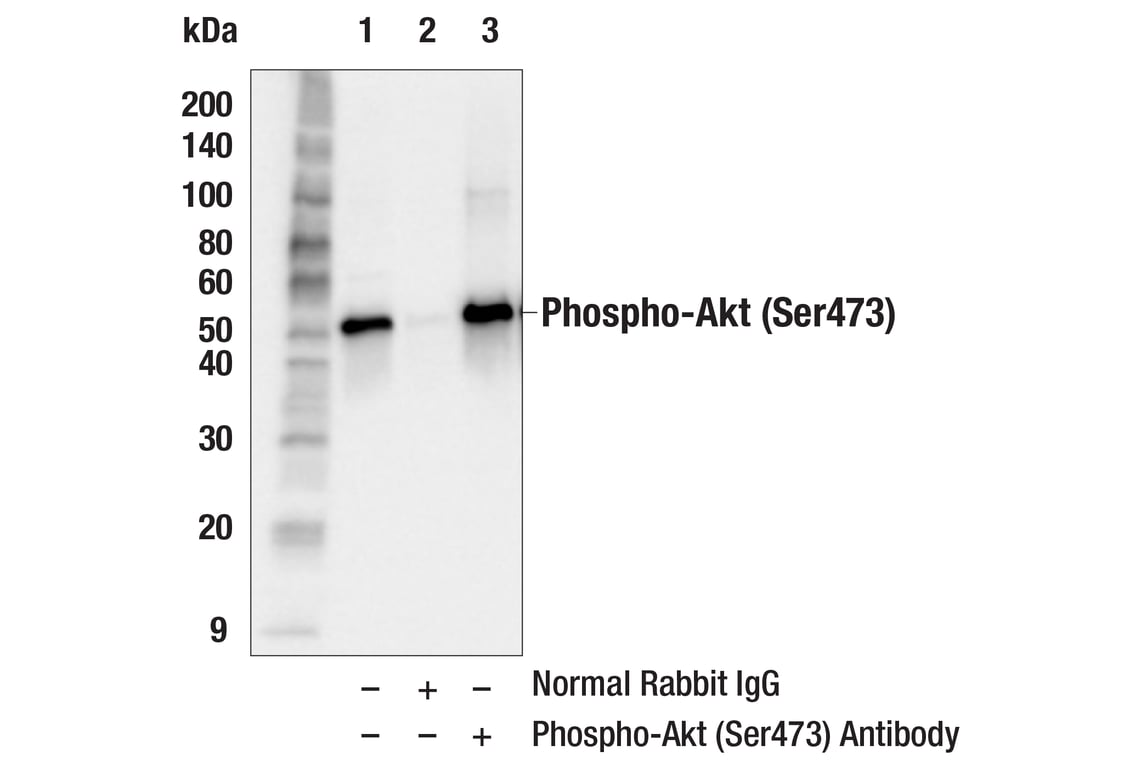
Immunoprecipitation of phospho-Akt (Ser473) from C2C12 cell extracts. Cells were treated with Insulin (100 ng/mL, 10 min.). Lane 1 is 10% input, lane 2 is Normal Rabbit IgG #2729, and lane 3 is Phospho-Akt (Ser473) Antibody. Western blot analysis was performed using Phospho-Akt (Ser473) (D9E) XP® Rabbit mAb #4060. Mouse Anti-rabbit IgG (Conformation Specific) (L27A9) mAb #3678 was used as a secondary antibody.
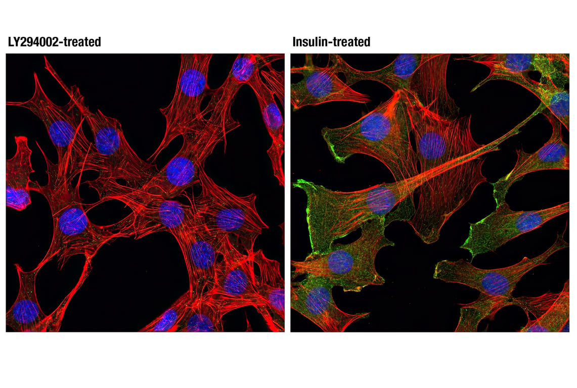
Confocal immunofluorescent analysis of C2C12 cells, treated with LY294002 #9901 (50 uM, 2 hrs; left) or insulin-treated (100 ng/mL, 30 min; right), using Phospho-Akt (Ser473) Antibody (green). Actin filaments were labeled with DY-554 phalloidin (red). Blue pseudocolor = DRAQ5® #4084 (fluorescent DNA dye).