


 全部商品分类
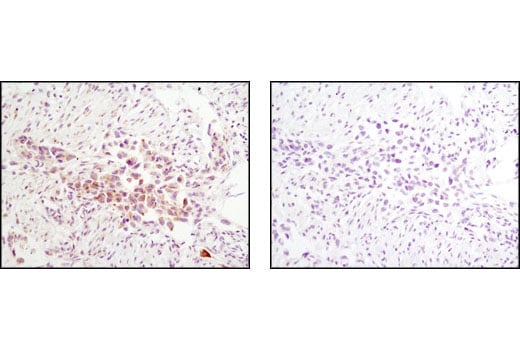


Phospho-Jak Family Antibody Sampler Kit

 下载产品说明书 下载SDS
 用小程序,查商品更便捷


 收藏
收藏
 对比
对比 咨询
咨询
The Phospho-Jak Family Antibody Sampler Kit provides an economical means of detecting the activation of Jak family members using phospho-specific and control antibodies. The kit includes enough antibody to perform two western blot experiments with each primary antibody.
参考图片
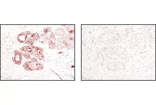
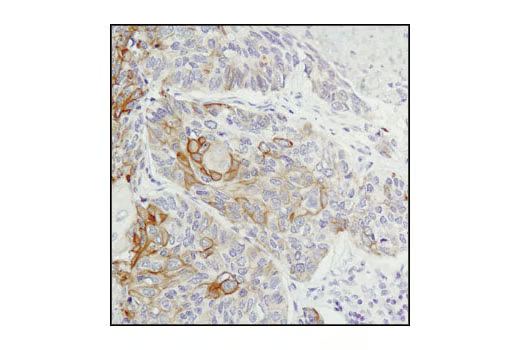
Immunohistochemical analysis of paraffin-embedded human lung carcinoma using Jak2 (D2E12) XP® Rabbit mA in the presence of control peptide (left) or Jak2 Blocking Peptide #1039 (right).
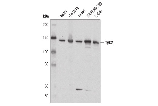
Western blot analysis of extracts from various cell lines using Tyk2 (D4I5T) Rabbit mAb.
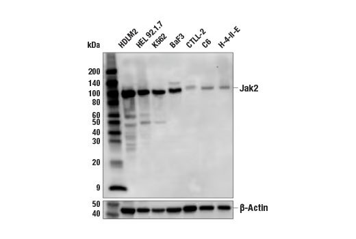
Western blot analysis of extracts from various cell lines using Jak2 (D2E12) XP® Rabbit mAb #3230 (upper) or β-Actin (D6A8) Rabbit mAb #8457 (lower).
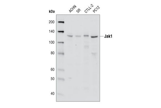
Western blot analysis of extracts from ACHN, SR, CTLL-2 and PC12 cell lines using Jak1 (6G4) Rabbit mAb.
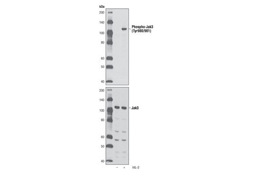
Western blot analysis of extracts from NK-92 cells, untreated or treated with Human Interleukin-2 (hIL-2) #8907 (10 ng/ml, 15 minutes), using Phospho-Jak3 (Tyr980/981) (D44E3) Rabbit mAb (upper) or Jak3 Antibody #3775 (lower).
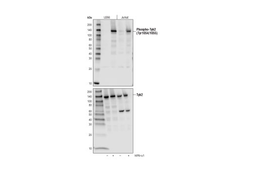
Western blot analysis of extracts from serum-starved U266 and Jurkat cells, untreated (-) or treated with Human Interferon-α1 (hIFN-α1) #8927 (50 ng/ml, 15 min; +) using Phospho-Tyk2 (Tyr1054/1055) (D7T8A) Rabbit mAb (upper) or total Tyk2 (D4I5T) Rabbit mAb #14193 (lower).
After the primary antibody is bound to the target protein, a complex with HRP-linked secondary antibody is formed. The LumiGLO® is added and emits light during enzyme catalyzed decomposition.
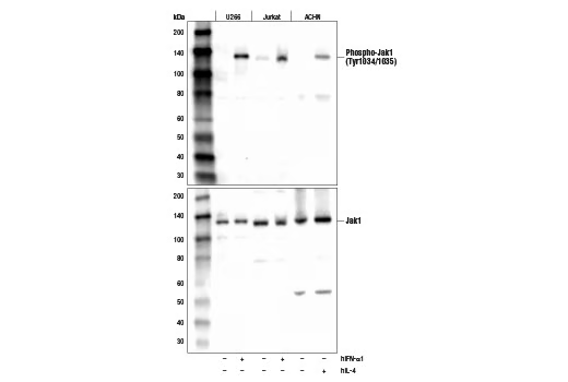
Western blot analysis of extracts from various cell lines, serum-starved overnight (-) followed by treatment with Human Interferon-α1 #8927 (hIFN-α1, 10 ng/ml, 15 min; +) or Human Interleukin-4 #8919 (hIL-4, 10 ng/ml, 10 min; +) using Phospho-Jak1 (Tyr1034/1035) (D7N4Z) Rabbit mAb (upper) or Jak1 (6G4) Rabbit mAb (lower).
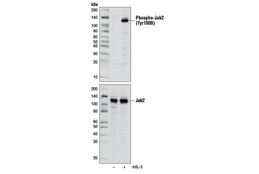
Western blot analysis of extracts from BaF3 cells, untreated or treated with Mouse Interleukin-3 (mIL-3) #8923 (10 ng/ml, 5 min), using Phospho-Jak2 (Tyr1008) (D4A8) Rabbit mAb (upper) or total Jak2 (D2E12) XP® Rabbit mAb #3230 (lower).
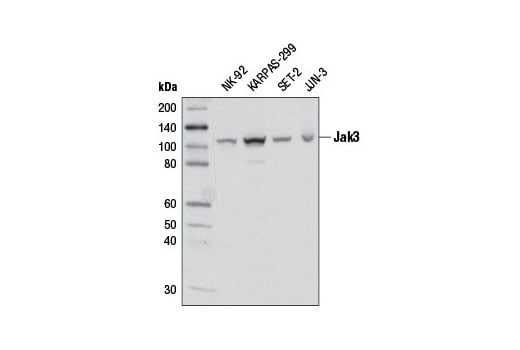
Western blot analysis of extracts from various cell lines using Jak3 (D1H3) Rabbit mAb.
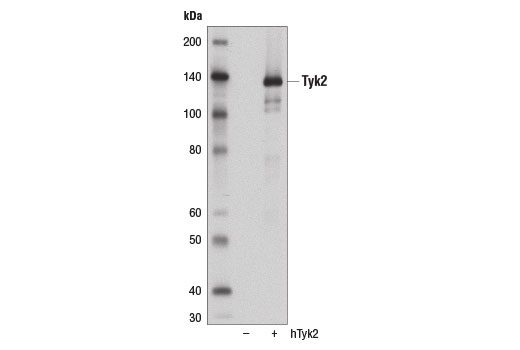
Western blot analysis of extracts from 293T cells, mock transfected (-) or transfected with a construct expressing full-length human Tyk2 (hTyk2; +), using Tyk2 (D4I5T) Rabbit mAb.
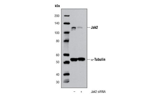
Western blot analysis of extracts from K-562 cells, transfected with 100 nM SignalSilence® Control siRNA (Unconjugated) #6568 (-) or Jak2 siRNA (+), using Jak2 (D2E12) XP® Rabbit mAb #3230 and α-Tubulin (11H10) Rabbit mAb #2125. The Jak2 (D2E12) XP® Rabbit mAb confirms silencing of Jak2 expression, while the α-Tubulin (11H10) Rabbit mAb is used to control for loading and specificity of Jak2 siRNA.
Immunohistochemical analysis of paraffin-embedded human breast carcinoma using Jak1 (6G4) Rabbit mAb #3344, in the presence of control peptide (left) or Jak1 Blocking Peptide (right).
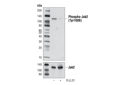
Western blot analysis of extracts from SET-2 cells, untreated or treated with the Jak2 inhibitor FLLL31 (10 μM, 4 hr), using Phospho-Jak2 (Tyr1008) (D4A8) Rabbit mAb (upper) or total Jak2 (D2E12) XP® Rabbit mAb #3230 (lower).
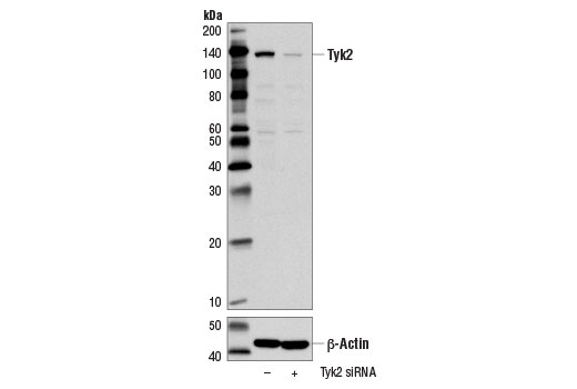
Western blot analysis of extracts from MCF7 cells, transfected with 100 nM SignalSilence® Control siRNA (Unconjugated) #6568 (-) or SignalSilence® Tyk2 siRNA #14254 (+), using Tyk2 (D4I5T) Rabbit mAb (upper) or β-Actin (D6A8) Rabbit mAb #8457 (lower). The Tyk2 (D4I5T) Rabbit mAb confirms silencing of Tyk2 expression, while the β-Actin (D6A8) Rabbit mAb is used as a loading control.
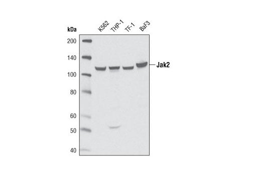
Western blot analysis of extracts from K-562, THP-1, TF-1 and BaF3 cell lines using Jak2 (D2E12) XP® Rabbit mAb.
Immunohistochemical analysis of paraffin-embedded human breast carcinoma using Jak1 (6G4) Rabbit mAb.
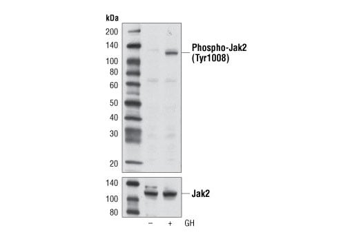
Western blot analysis of extracts from LNCaP cells, untreated or treated with growth hormone (GH) (100 ng/ml, 5 min), using Phospho-Jak2 (Tyr1008) (D4A8) Rabbit mAb (upper) or total Jak2 (D2E12) XP® Rabbit mAb #3230 (lower).
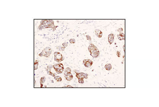
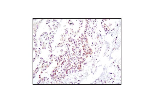
Immunohistochemical analysis of paraffin-embedded human lung carcinoma using Jak2 (D2E12) XP® Rabbit mAb.
Immunohistochemical analysis of paraffin-embedded human lung carcinoma using Jak1 (6G4) Rabbit mAb.