


 全部商品分类


Phospho-MAPK Family Antibody Sampler Kit

 下载产品说明书 下载SDS
 用小程序,查商品更便捷


 收藏
收藏
 对比
对比 咨询
咨询
The Phospho-MAPK Family Antibody Sampler Kit provides an economical means of evaluating the phosphorylation state of p38, p44/42, and SAPK/JNK mitogen-activated protein kinases. The kit contains enough primary and secondary antibodies to perform two western blot experiments.
参考图片
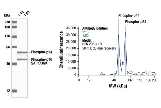
Simple Western™ analysis of lysates (1.0 mg/mL) from HEK 293 cells treated with UV (50 mJ, 30 min recovery) using Phospho-SAPK/JNK (Thr183/Tyr185) (81E11) Rabbit mAb #4668. The virtual lane view (left) shows two target bands (as indicated) at 1:10 and 1:50 dilutions of primary antibody. The corresponding electropherogram view (right) plots chemiluminescence by molecular weight along the capillary at 1:10 (blue line) and 1:50 (green line) dilutions of primary antibody. This experiment was performed under reducing conditions on the Jess™ Simple Western instrument from ProteinSimple, a BioTechne brand, using the 12-230 kDa separation module.
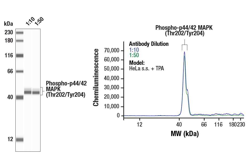
Simple Western™ analysis of lysates (0.1 mg/mL) from serum-starved HeLa cells treated with TPA (400 nM, 4 hours) using Phospho-p44/42 MAPK (Erk1/2) (Thr202/Tyr204) (D13.14.4E) XP® Rabbit mAb #4370. The virtual lane view (left) shows the target bands (as indicated) at 1:10 and 1:50 dilutions of primary antibody. The corresponding electropherogram view (right) plots chemiluminescence by molecular weight along the capillary at 1:10 (blue line) and 1:50 (green line) dilutions of primary antibody. This experiment was performed under reducing conditions on the Jess™ Simple Western instrument from ProteinSimple, a BioTechne brand, using the 12-230 kDa separation module.
Immunohistochemical analysis of paraffin-embedded human urothelial carcinoma using Phospho-p44/42 MAPK (Erk1/2) (Thr202/Tyr204) (D13.14.4E) XP® Rabbit mAb performed on the Leica BOND RX.
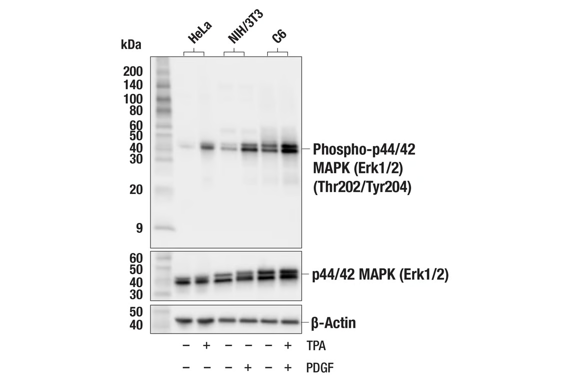
Western blot analysis of extracts from various cell lines treated with TPA (200nM, 4 hr) or PDGF (100ng/mL, 10 min) as indicated, using Phospho-p44/42 MAPK (Erk1/2) (Thr202/Tyr204) (D13.14.4E) XP® Rabbit mAb (upper), p44/42 MAPK (Erk1/2) (137F5) Rabbit mAb #4695 (middle), or β-Actin (D6A8) Rabbit mAb #8457 (lower).
Western blot analysis of extracts from Jurkat cells, treated with U0126 (10 µM, 1 hour) (negative control) or treated with TPA (200 nM, 20 min) (positive control), using p44/42 MAPK (Erk1/2) (L34F12) Mouse mAb #4696 (Panel A) and Phospho-p44/42 MAPK (Erk1/2) (Thr202/Tyr204) (D13.14.4E) XP® Rabbit mAb #4370 (Panel B). Anti-mouse IgG (H+L) (DyLight 680 Conjugate) #5470 (red) and Anti-rabbit IgG (H+L) (DyLight 800 4X PEG Conjugate) #5151 (green) were used as secondary antibodies.
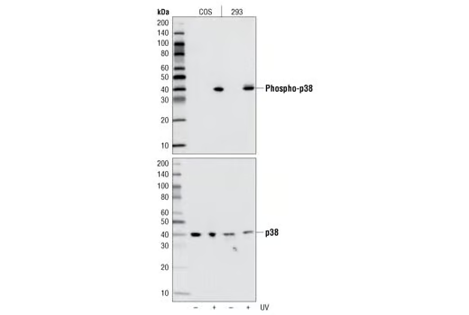
Western blot analysis of extracts from COS and 293 cells, untreated or UV-treated, using Phospho-p38 MAPK (Thr180/Tyr182) (D3F9) XP® Rabbit mAb (upper) or p38 MAPK Antibody #9212 (lower).
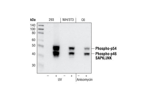
Western blot analysis of extracts from 293 cells, untreated or UV-treated, NIH/3T3 cells, untreated or UV-treated and C6 cells, untreated or anisomycin-treated, using Phospho-SAPK/JNK (Thr183/Tyr185) (81E11) Rabbit mAb.
After the primary antibody is bound to the target protein, a complex with HRP-linked secondary antibody is formed. The LumiGLO® is added and emits light during enzyme catalyzed decomposition.
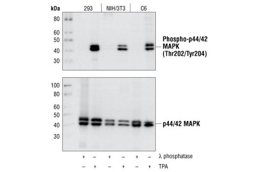
Western blot analysis of extracts from 293, NIH/3T3 and C6 cells, treated with λ phosphatase or TPA #4174 as indicated, using Phospho-p44/42 MAPK (Erk1/2) (Thr202/Tyr204) (D13.14.4E) XP® Rabbit mAb (upper), or p44/42 MAPK (Erk1/2) (137F5) Rabbit mAb #4695 (lower).
Immunohistochemical analysis of paraffin-embedded human squamous cell carcinoma of the skin using Phospho-p44/42 MAPK (Erk1/2) (Thr202/Tyr204) (D13.14.4E) XP® Rabbit mAb performed on the Leica BOND RX.
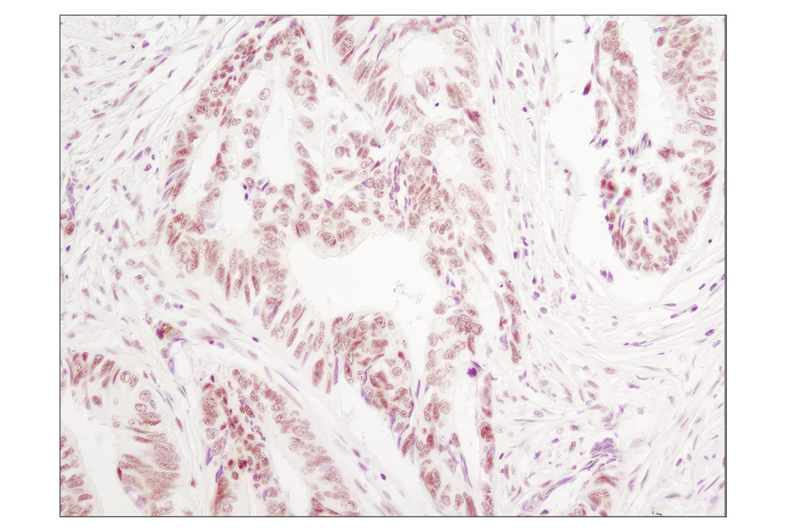
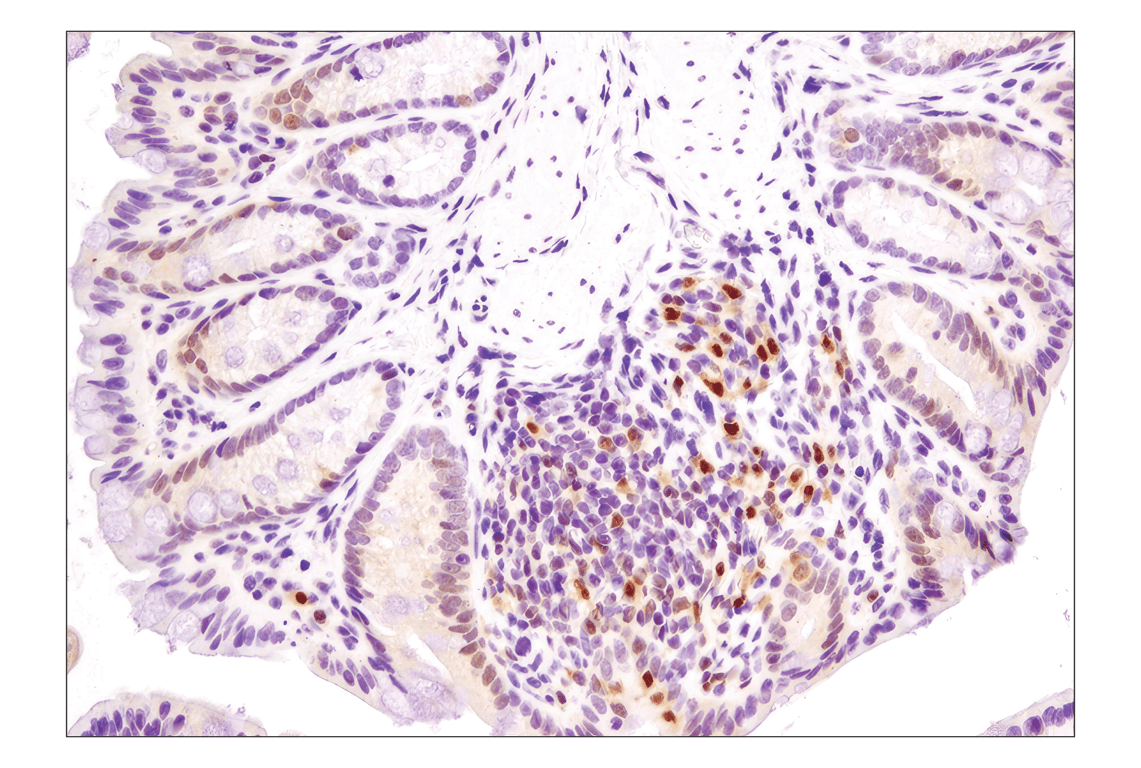
Immunohistochemical analysis of paraffin-embedded human colon carcinoma using Phospho-p38 MAPK (Thr180/Tyr182) (D3F9) XP® Rabbit mAb.
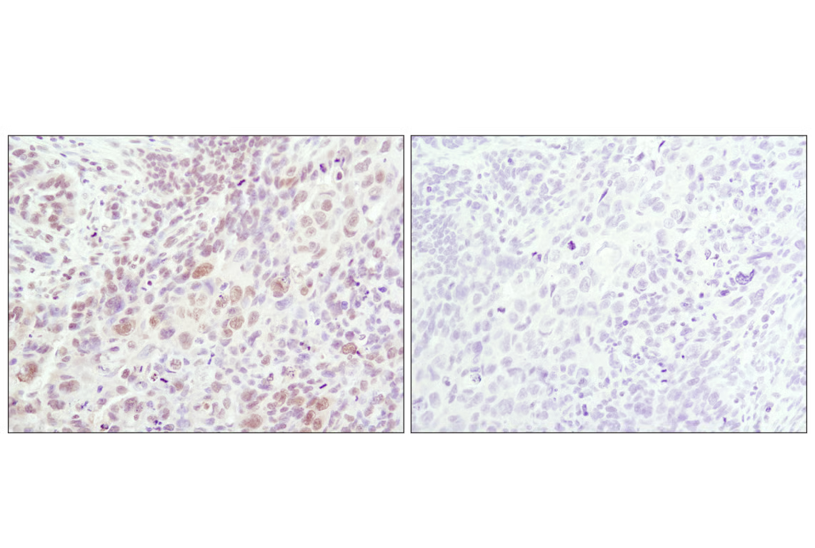
Immunohistochemical analysis of paraffin-embedded human lung carcinoma using Phospho-SAPK/JNK (Thr183/Tyr185) (81E11) Rabbit mAb in the presence of control peptide (left) or Phospho-SAPK/JNK (Thr183/Tyr185) Blocking Peptide #1215 (right).
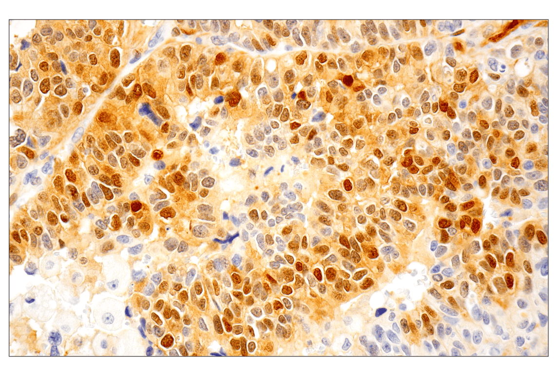
Immunohistochemical analysis of paraffin-embedded human breast carcinoma using Phospho-p44/42 MAPK (Erk1/2) (Thr202/Tyr204) (D13.14.4E) XP® Rabbit mAb.
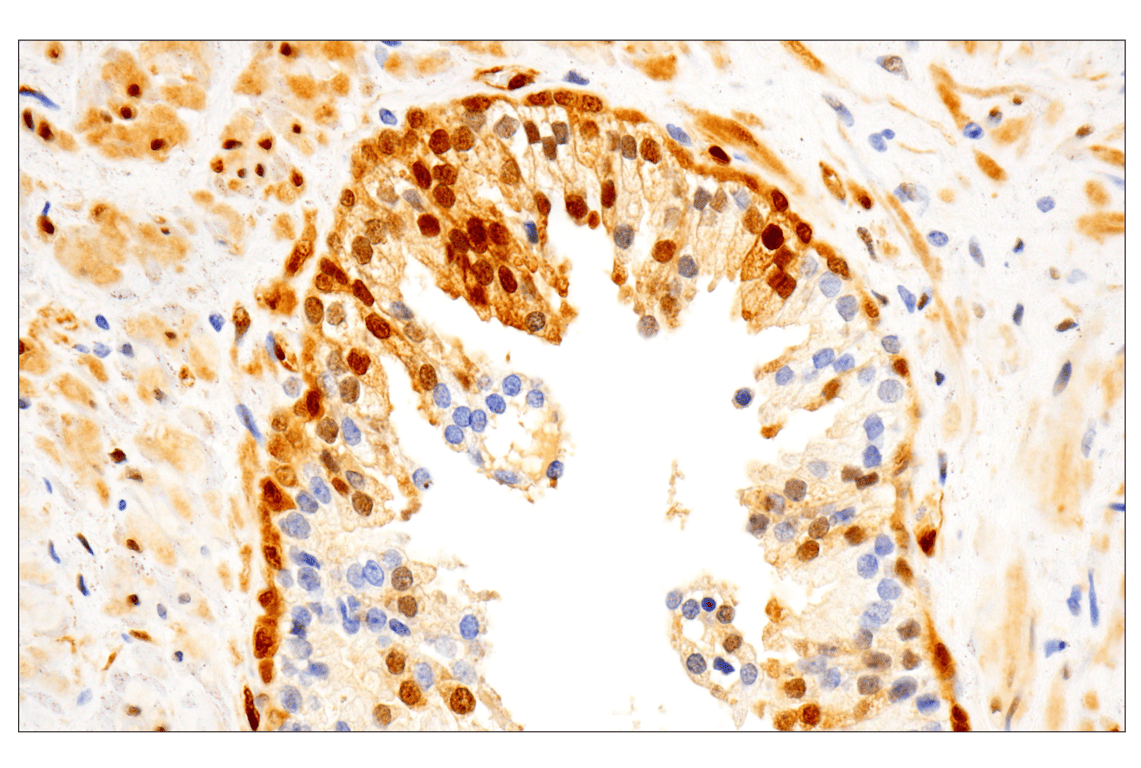
Immunohistochemical analysis of paraffin-embedded human endometrioid adenocarcinoma using Phospho-p44/42 MAPK (Erk1/2) (Thr202/Tyr204) (D13.14.4E) XP® Rabbit mAb performed on the Leica BOND RX.
Immunohistochemical analysis of paraffin-embedded mouse colon using Phospho-p38 MAPK (Thr180/Tyr182) (D3F9) XP® Rabbit mAb.
Immunohistochemical analysis of paraffin-embedded 293T cells untreated (left) or UV-treated (right) using Phospho-SAPK/JNK (Thr183/Tyr185) (81E11) Rabbit mAb.
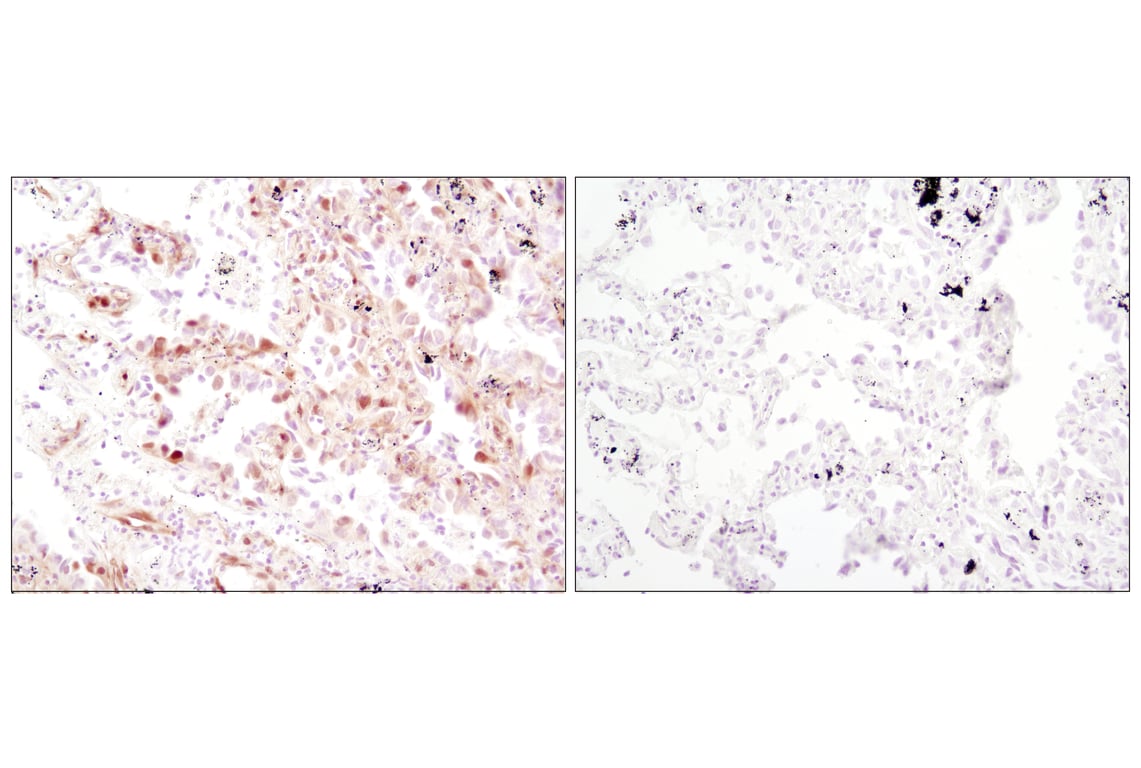
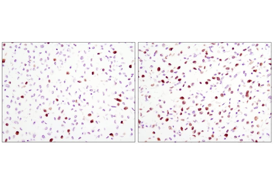
Immunohistochemical analysis of paraffin-embedded human lung carcinoma, untreated (left) or λ phosphatase-treated (right), using Phospho-p44/42 MAPK (Erk1/2) (Thr202/Tyr204) (D13.14.4E) XP® Rabbit mAb.
Immunohistochemical analysis of paraffin-embedded human prostate adenocarcinoma using Phospho-p44/42 MAPK (Erk1/2) (Thr202/Tyr204) (D13.14.4E) XP® Rabbit mAb performed on the Leica BOND RX.
Immunohistochemical analysis of paraffin-embedded 293T cell pellets, untreated (left) or UV-treated (right), using Phospho-p38 MAPK (Thr180/Tyr182) (D3F9) XP® Rabbit mAb.
Immunohistochemical analysis using Phospho-p44/42 MAPK (Erk1/2) (Thr202/Tyr204) (D13.14.4E) XP® Rabbit mAb on SignalSlide™ Phospho-p44/42 MAPK (Thr202/Tyr204) IHC Controls #8103 (paraffin-embedded NIH/3T3 cells, treated with U0126 #9903 (left) or TPA #4174 (right).
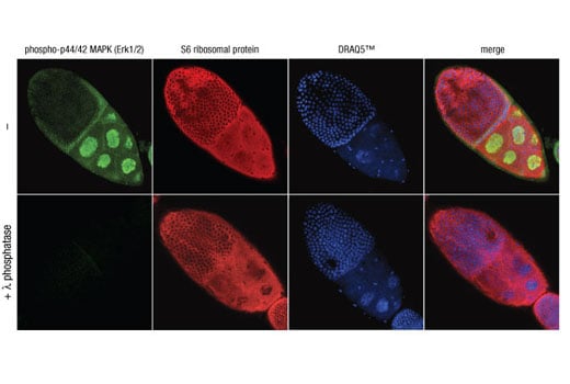
Confocal immunofluorescent analysis of Drosophila egg chambers, untreated (top) or λ phosphatase-treated (bottom), using Phospho-p44/42 MAPK (Erk1/2) (Thr202/Tyr204) (D13.14.4E) XP® Rabbit mAb #4370 (green) and S6 Ribosomal Protein (54D2) Mouse mAb #2317 (red). Blue pseudocolor = DRAQ5® #4084 (fluorescent DNA dye).
Confocal immunofluorescent analysis of COS cells, untreated (left) or anisomycin-treated (right) using Phospho-p38 MAPK (Thr180/Tyr182) (D3F9) XP® Rabbit mAb (green). Actin filaments have been labeled with DY-554 phalloidin (red).
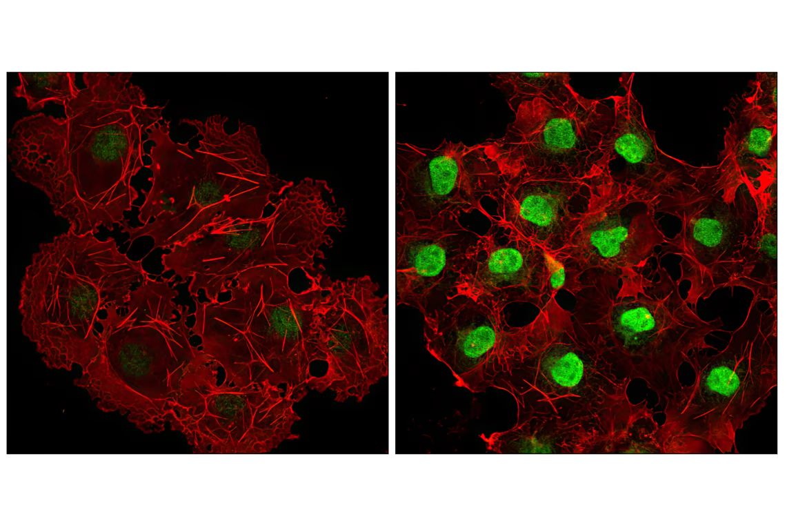
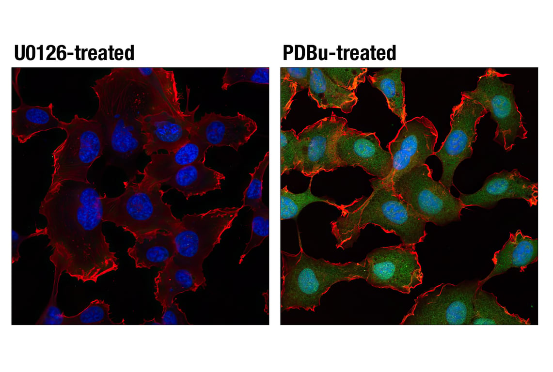
Confocal immunofluorescent analysis of HT1080 cells, starved overnight then treated with U0126 #9903 (10 uM, 2 h; left) or PDBu (Phorbol 12,13-Dibutyrate) #12808 (100 nM, 15 m; right) using Phospho-p44/42 MAPK (Erk1/2) (Thr202/Tyr204) (D13.14.4E) XP® Rabbit mAb #4370 (green) and β-Actin (8H10D10) Mouse mAb #3700 (red). Blue pseudocolor = DRAQ5® #4084 (fluorescent DNA dye).
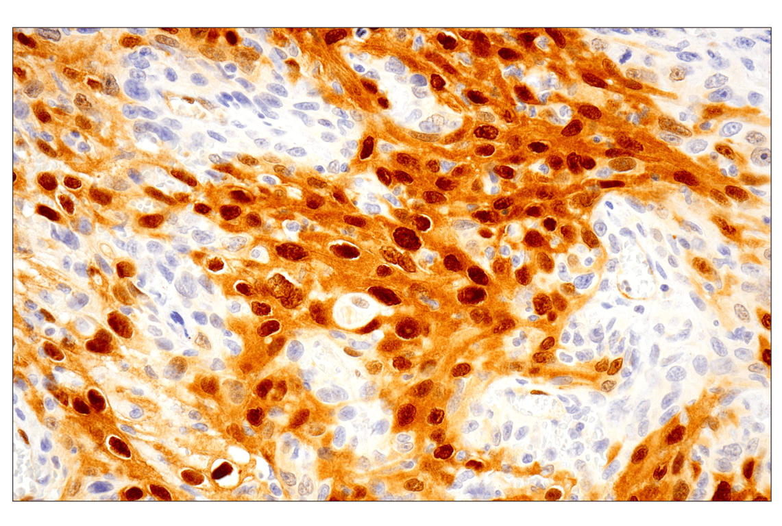
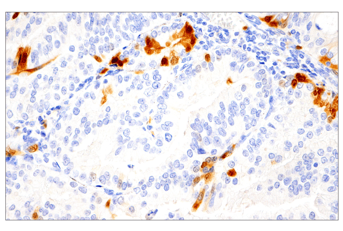
Immunohistochemical analysis of paraffin-embedded human lung adenocarcinoma using Phospho-p44/42 MAPK (Erk1/2) (Thr202/Tyr204) (D13.14.4E) XP® Rabbit mAb.
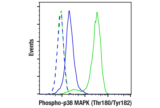
Flow cytometric analysis of Jurkat cells, untreated (blue) or treated with Anisomycin (25µM, 30 min; green) using Phospho-p38 MAPK (Thr180/Tyr182) (D3F9) XP® Rabbit mAb (solid lines) or concentration-matched Rabbit (DA1E) mAb IgG XP® Isotype Control #3900 (dashed lines). Anti-rabbit IgG (H+L), F(ab')2 Fragment (Alexa Fluor® 488 Conjugate) #4412 was used as a secondary antibody.
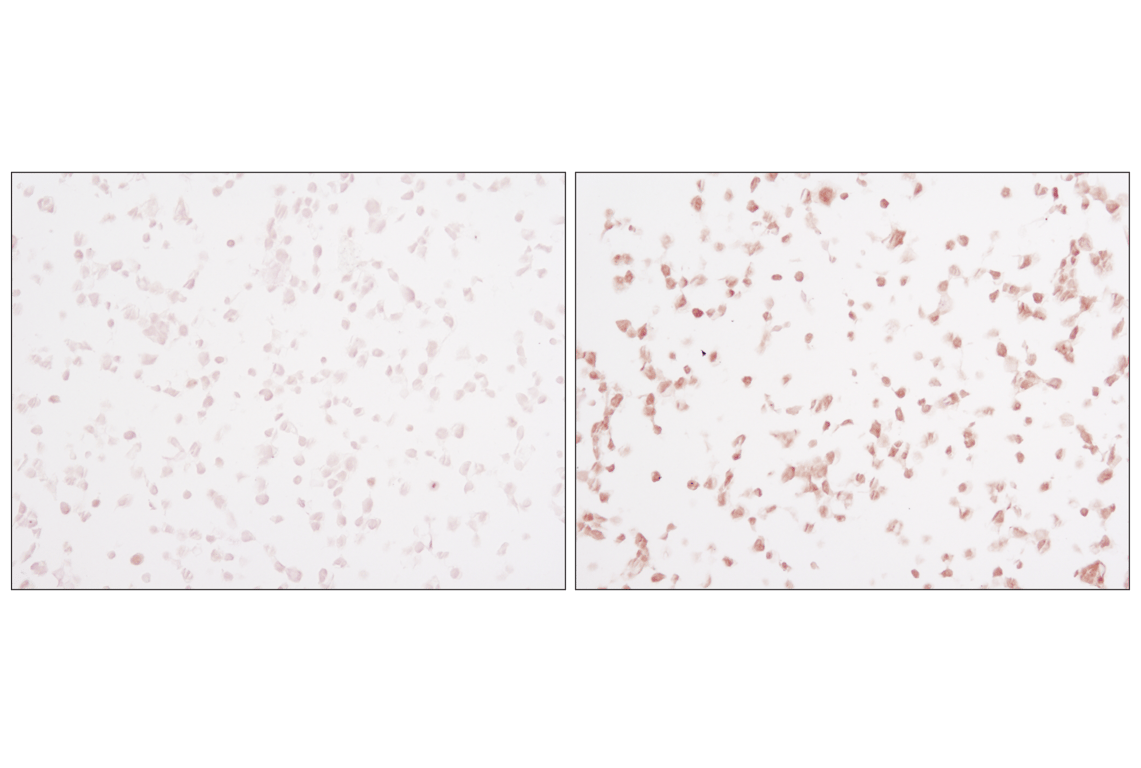
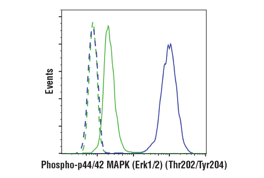
Flow cytometric analysis of Jurkat cells, treated with U0126 (10 µM, 2 hrs; green) or treated with TPA #4174 (200 nM, 30 min; blue) using Phospho-p44/42 MAPK (Erk1/2) (Thr202/Tyr204) (D13.14.4E) XP® Rabbit mAb (solid lines) or concentration-matched Rabbit (DA1E) mAb IgG XP® Isotype Control #3900 (dashed lines). Anti-rabbit IgG (H+L), F(ab')2 Fragment (Alexa Fluor® 488 Conjugate) #4412 was used as a secondary antibody.
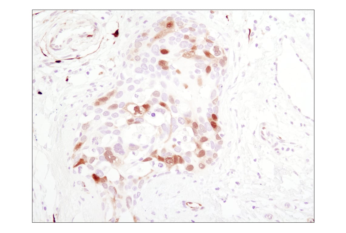
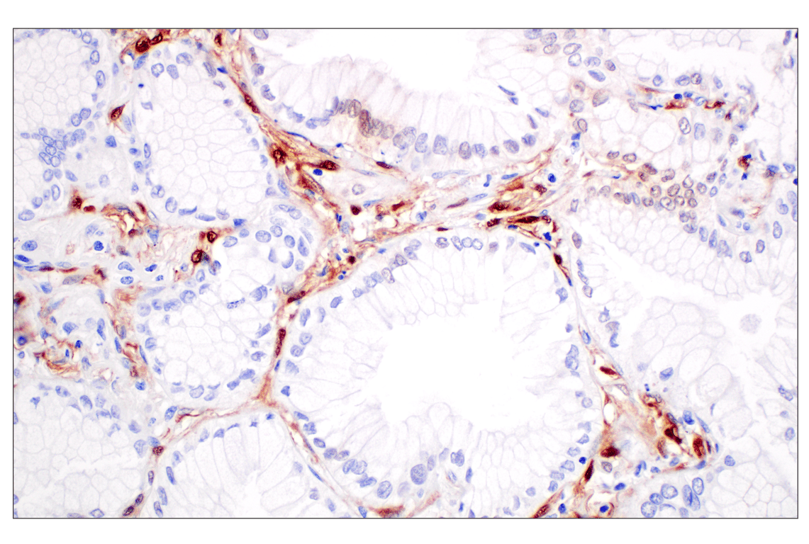
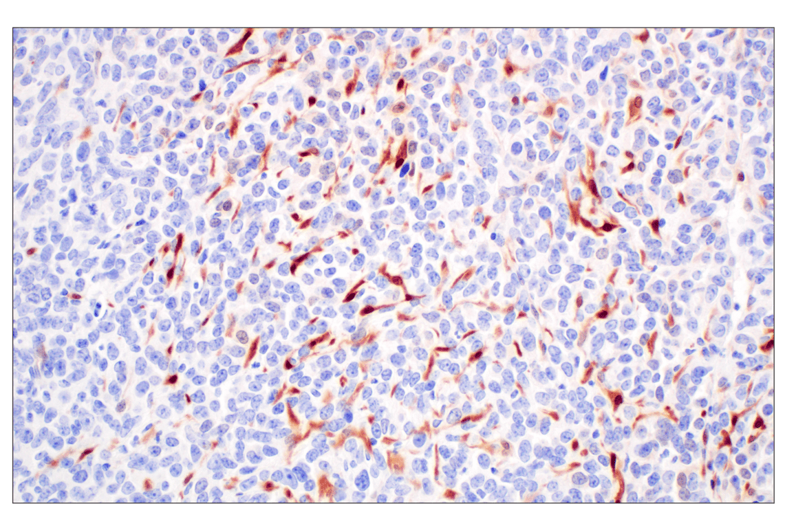
Immunohistochemical analysis of paraffin-embedded 4T1 syngeneic tumor using Phospho-p44/42 MAPK (Erk1/2) (Thr202/Tyr204) (D13.14.4E) XP® Rabbit mAb.
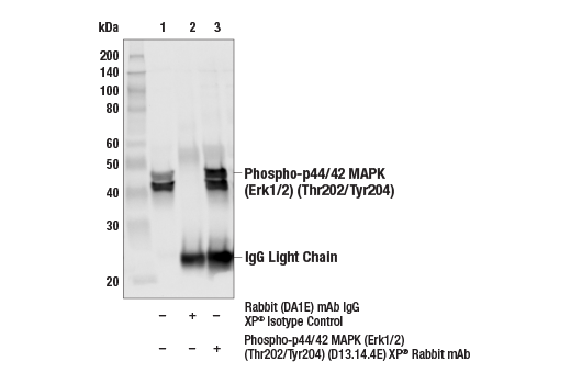
Immunoprecipitation of Phospho-p44/42 MAPK (Erk1/2) (Thr202/Tyr204) from 3T3 cell extracts. Cells were treated with TPA, (200 nM, 15 min). Lane 1 is 10% input, lane 2 is Rabbit (DA1E) mAb IgG XP® Isotype Control #3900, and lane 3 is Phospho-p44/42 MAPK (Erk1/2) (Thr202/Tyr204) (D13.14.4E) XP® Rabbit mAb. Western blot was performed using Phosphop44/42 MAPK (Erk1/2) (Thr202/Tyr204) (D13.14.4E) XP® Rabbit mAb. Mouse Anti-rabbit IgG (Light-Chain Specific) (D4W3E) mAb #45262 was used as a secondary antibody.