


 下载产品说明书
 用小程序,查商品更便捷


 收藏
收藏
 对比
对比 咨询
咨询
Specificity/Sensitivity
参考图片
Flow cytometric analysis of HeLa cells, untreated (blue) or UV-treated (green), using Phospho-MAPKAPK-2 (Thr334) (27B7) Rabbit mAb.
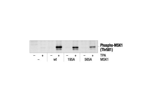
Western blot analysis of extracts from 293 cells, transfected with wt MSK1 or mutant MSK1, untreated or TPA-treated (200 nM), using Phospho-MSK1 (Thr581) Antibody.
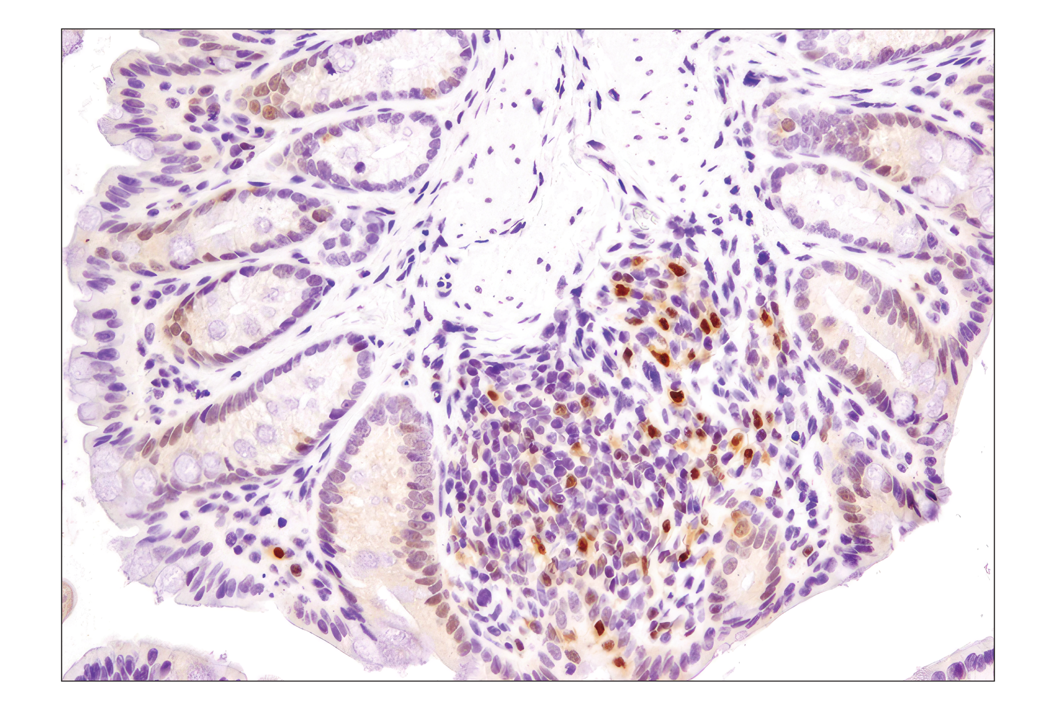
Immunohistochemical analysis of paraffin-embedded human breast carcinoma, showing nuclear localization, using Phospho-MSK1 (Thr581) Antibody.
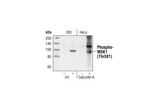
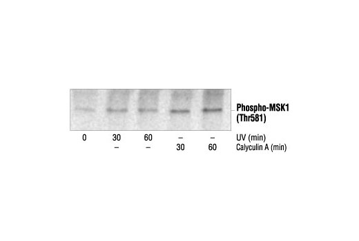
Western blot analysis of extracts from 293 cells, untreated, UV-treated or calyculin A-treated, using Phospho-MSK1 (Thr581) Antibody.
After the primary antibody is bound to the target protein, a complex with HRP-linked secondary antibody is formed. The LumiGLO* is added and emits light during enzyme catalyzed decomposition.
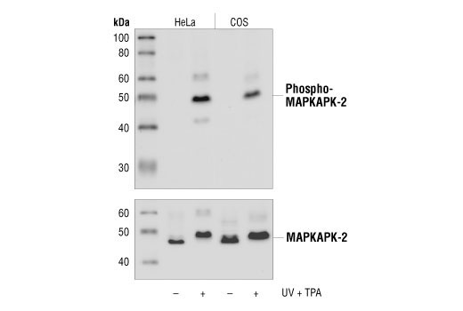
Western blot analysis of extracts from untreated or UV+TPA-treated HeLa and COS cells, using Phospho-MAPKAPK-2 (Thr334) (27B7) Rabbit mAb (upper), or MAPKAPK-2 Antibody #3042 (lower).
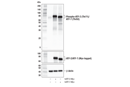
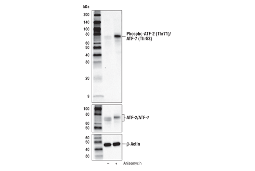
Western blot analysis of extracts from untreated or UV-treated COS cells, NIH/3T3 cells and C6 cells, using Phospho-ATF-2 (Thr71) (11G2) Rabbit mAb (upper), or ATF-2 (20F1) Rabbit mAb #9226 (lower).
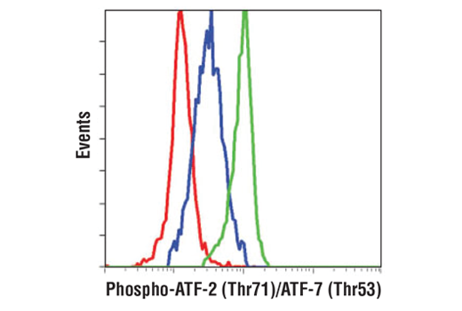
Flow cytometric analysis of THP-1 cells, untreated (blue) or Anisomycin treated (green), using Phospho-ATF-2 (Thr71) (11G2) Rabbit mAb compared to a nonspecific negative control antibody (red).
Western blot analysis of extracts from 293 cells untreated or treated with UV and HeLa cells untreated or treated with calyculin A, using Phospho-MSK1 (Thr581) Antibody.
Confocal immunofluorescent analysis of HeLa cells, untreated (left), UV-treated (center) or UV and lambda phosphatase-treated (right), using Phospho-MAPKAPK-2 (Thr334) (27B7) Rabbit mAb (green). Actin filaments have been labeled with DY-554 phalloidin (red).
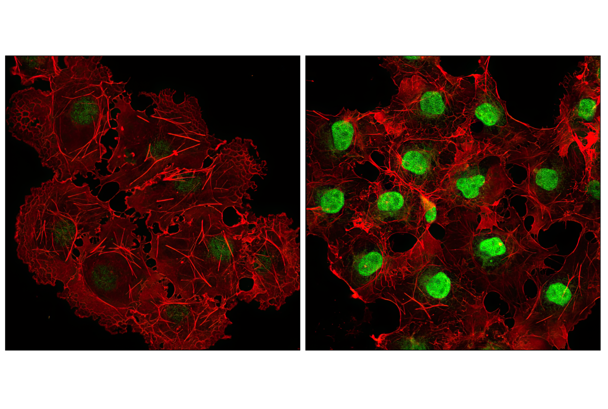
Confocal immunofluorescent analysis of COS cells, untreated (left) or anisomycin-treated (right) using Phospho-p38 MAPK (Thr180/Tyr182) (D3F9) XP® Rabbit mAb (green). Actin filaments have been labeled with DY-554 phalloidin (red).
Western blot analysis of extracts from 293 cells untreated or treated with UV and HeLa cells untreated or treated with calyculin A, using Phospho-MSK1 (Thr581) Antibody #9595.
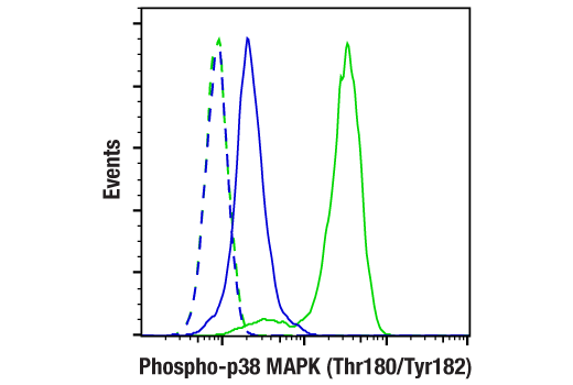
Flow cytometric analysis of Jurkat cells, untreated (blue) or anisomycin-treated (green), using Phospho-p38 MAPK (Thr180/Tyr182) (D3F9) XP® Rabbit mAb compared to a nonspecific negative control antibody (red).
Western blot analysis of extracts from untreated or UV-treated COS, NIH/3T3 and C6 cells, using Phospho-MKK3/6 (Ser189/207) (22A8) Rabbit mAb #9236.
Western blot analysis of extracts from untreated or UV-treated COS cells, NIH/3T3 cells and C6 cells, using Phospho-ATF-2 (Thr71) (11G2) Rabbit mAb #5112 (upper), or ATF-2 (20F1) Rabbit mAb #9226 (lower).
Western blot analysis of extracts from untreated or UV+TPA-treated HeLa and COS cells, using Phospho-MAPKAPK-2 (Thr334) (27B7) Rabbit mAb #3007 (upper), or MAPKAPK-2 Antibody #3042 (lower).
Immunohistochemical analysis of paraffin-embedded 293T cell pellets, untreated (left) or UV-treated (right), using Phospho-p38 MAPK (Thr180/Tyr182) (D3F9) XP® Rabbit mAb.
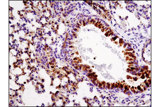
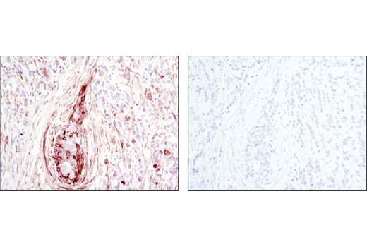
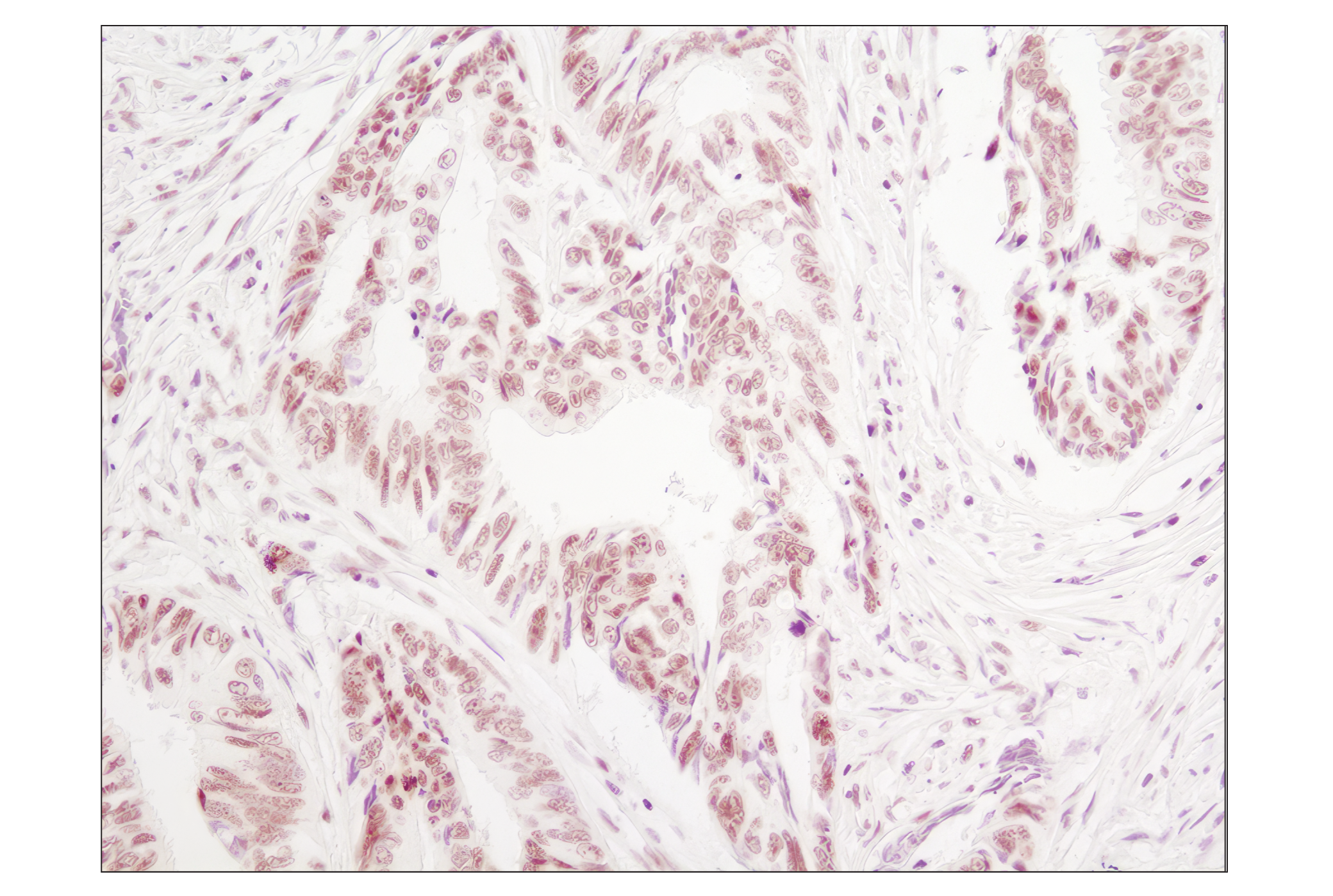
Immunohistochemical analysis of paraffin-embedded human colon carcinoma using Phospho-p38 MAPK (Thr180/Tyr182) (D3F9) XP® Rabbit mAb.
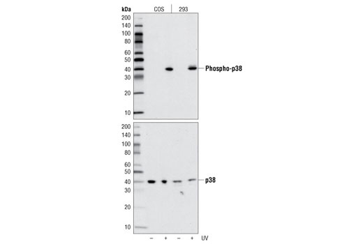
Western blot analysis of extracts from COS and 293 cells, untreated or UV-treated, using Phospho-p38 MAPK (Thr180/Tyr182) (D3F9) XP® Rabbit mAb (upper) or p38 MAPK Antibody #9212 (lower).
Western blot analysis of extracts from COS and 293 cells, untreated or UV-treated, using Phospho-p38 MAPK (Thr180/Tyr182) (D3F9) Rabbit mAb #4511 (upper) or p38 MAPK Antibody #9212 (lower).
Immunohistochemical analysis of paraffin-embedded HeLa cell pellets, control (left) or UV-treated (right), using Phospho-HSP27 (Ser82) (D1H2) XP® Rabbit mAb.
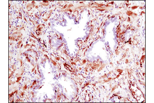
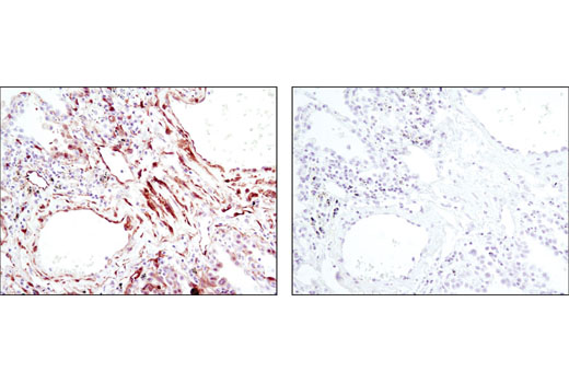
Immunohistochemical analysis of paraffin-embedded human prostate carcinoma using Phospho-HSP27 (Ser82) (D1H2) XP® Rabbit mAb.
Immunohistochemical analysis of paraffin-embedded human breast carcinoma, control (left) or λ phosphatase-treated (right), using Phospho-HSP27 (Ser82) (D1H2) XP® Rabbit mAb.
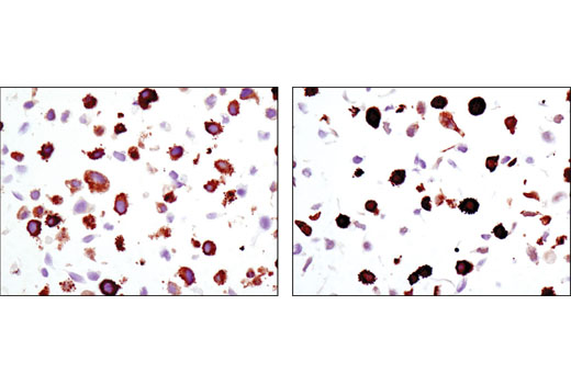
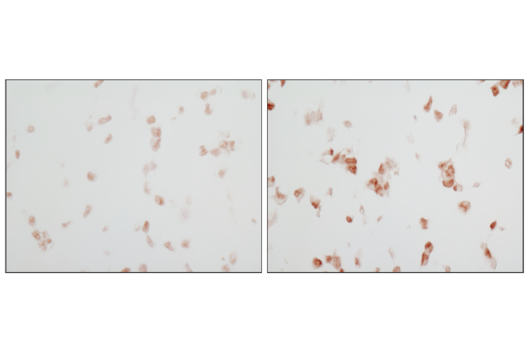
Immunohistochemical analysis of paraffin-embedded human lung carcinoma using Phospho-HSP27 (Ser82) (D1H2) XP® Rabbit mAb in the presence of control peptide (left) or antigen-specific peptide (right).
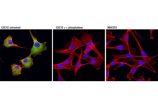
Confocal immunofluorescent analysis of C2C12 cells, untreated (left) or treated with λ phosphatase (middle), and NIH/3T3 cells (right) using Phospho-HSP27 (Ser82) (D1H2) XP® Rabbit mAb (green). Actin filaments were labeled with DY-554 phalloidin (red). Blue pseudocolor = DRAQ5® #4084 (fluorescent DNA dye). Negative staining in NIH/3T3 cells is in agreement with the observation that NIH/3T3 cells do not express HSP27 under basal conditions (5,7).
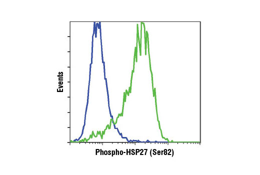
Flow cytometric analysis of HeLa cells, untreated (blue) or UV-treated (green), using Phospho-HSP27 (Ser82) (D1H2) XP® Rabbit mAb.
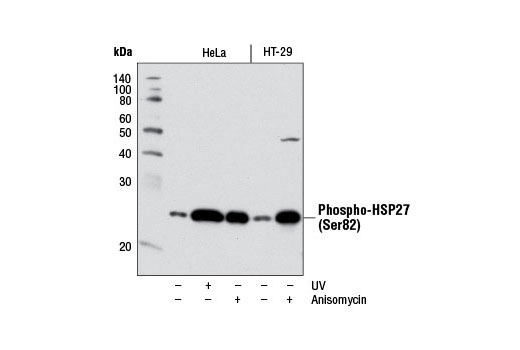
Western blot analysis of extracts from HeLa or HT-29 cells, untreated (-) or treated (+) with either UV (40 mJ/cm2 with 30 min recovery) or anisomycin (25 μg/mL, 30 min), using Phospho-HSP27 (Ser82) (D1H2) XP® Rabbit mAb.
Immunohistochemical analysis of paraffin-embedded mouse lung using Phospho-HSP27 (Ser82) (D1H2) XP® Rabbit mAb.
Western blot analysis of extracts from HeLa or HT-29 cells, untreated (-) or treated (+) with either UV (40 mJ/cm2 with 30 min recovery) or anisomycin (25 μg/mL, 30 min), using Phospho-HSP27 (Ser82) (D1H2) XP® Rabbit mAb #9709.
Confocal immunofluorescent analysis of HeLa cells, untreated (left) or UV-treated (40 mJ/cm2 with 30 min recovery; right), using Phospho-MKK3 (Ser189)/MKK6 (Ser207) (D8E9) Rabbit mAb (green). Actin filaments were labeled with DY-554 phalloidin (red). Blue pseudocolor = DRAQ5® #4084 (fluorescent DNA dye).
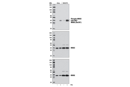
Western blot analysis of extracts from HeLa and NIH/3T3 cells, untreated (-) or treated with TPA #4174 (200 nM, 15 min; +), using Phospho-MKK3 (Ser189)/MKK6 (Ser207) (D8E9) Rabbit mAb (upper), MKK3 (D4C3) Rabbit mAb #8535 (middle), or MKK6 (D31D1) Rabbit mAb #8550 (lower).
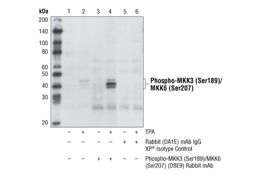
Immunoprecipitation of phospho-MKK3 (Ser189)/MKK6 (Ser207) from HeLa cells, untreated or treated with TPA #4174 (200 nM, 15 min), using Phospho-MKK3 (Ser189)/MKK6 (Ser207) (D8E9) Rabbit mAb (lanes 3 and 4) or Rabbit (DA1E) mAb IgG XP® Isotype Control #3900 (lanes 5 and 6). Lanes 1 and 2 are 10% input. Western blot analysis was performed using
Phospho-MKK3 (Ser189)/MKK6 (Ser207) Antibody #9231. Mouse Anti-rabbit IgG (Conformation Specific) (L27A9) mAb #3678 was used as a secondary antibody.

 危险品化学品经营许可证(不带存储) 许可证编号:沪(杨)应急管危经许[2022]202944(QY)
  营业执照(三证合一)