


 全部商品分类
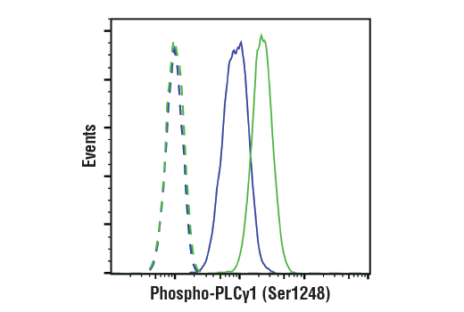


 用小程序,查商品更便捷


Monoclonal antibody is produced by immunizing animals with a synthetic peptide corresponding to residues surrounding Ser1248 of human PLCγ1 protein.

Product Usage Information
| Application | Dilution |
|---|---|
| Western Blotting | 1:1000 |
| Simple Western™ | 1:10 - 1:50 |
| Immunoprecipitation | 1:50 |
| Immunohistochemistry (Paraffin) | 1:100 - 1:400 |
| Immunofluorescence (Immunocytochemistry) | 1:100 - 1:200 |
| Flow Cytometry (Fixed/Permeabilized) | 1:800 |
Specificity/Sensitivity
Species Reactivity:
Human, Mouse, Monkey
Supplied in 10 mM sodium HEPES (pH 7.5), 150 mM NaCl, 100 µg/ml BSA, 50% glycerol and less than 0.02% sodium azide. Store at –20°C. Do not aliquot the antibody.
参考图片
Flow cytometric analysis of Jurkat cells, treated with U0126 #9903 (10uM, 2 hr; blue) or TPA #4174 (200nM, 30 min; green) using Phospho-PLCγ1 (Ser1248) (D25A9) Rabbit mAb (solid lines) or concentration-matched Rabbit (DA1E) mAb IgG XP® Isotype Control #3900 (dashed lines). Anti-rabbit IgG (H+L), F(ab')2 Fragment (Alexa Fluor® 488 Conjugate) was used as a secondary antibody.
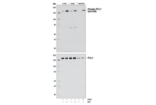
Western blot analysis of extracts from serum-starved A-431 and A549 cells, untreated (-) or treated (+) with hEGF #8916 (100 ng/mL, 15 min) or serum-starved NIH/3T3 cells, untreated (-) or treated (+) with hPDGF-BB #8912 (50 ng/mL, 15 min), using Phospho-PLCγ1 (Ser1248) (D25A9) Rabbit mAb (upper) or PLCγ1 (D9H10) XP® Rabbit mAb #5690 (lower).
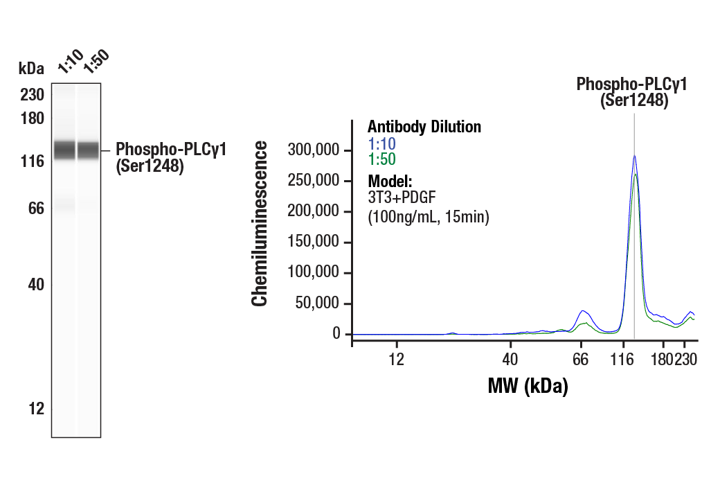
Simple Western™ analysis of lysates (0.8 mg/mL) from 3T3+PDGF (100ng/mL, 15min) cells using Phospho-PLCγ1 (Ser1248) (D25A9) Rabbit mAb #8713. The virtual lane view (left) shows the target band (as indicated) at 1:10 and 1:50 dilutions of primary antibody. The corresponding electropherogram view (right) plots chemiluminescence by molecular weight along the capillary at 1:10 (blue line) and 1:50 (green line) dilutions of primary antibody. This experiment was performed under reducing conditions on the Jess™ Simple Western instrument from ProteinSimple, a BioTechne brand, using the 12-230 kDa separation module.
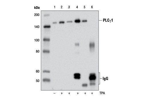
Immunoprecipitation (IP)/Western blot analysis of extracts from serum-starved HeLa cells, untreated (-) or treated (+) with TPA #4174 (100 nM, 15 min) prior to lysis in SDS (lanes 1 and 2) or IP lysis buffer (lane 3, TPA-treated only). IP Lysates were then subjected to immunoprecipitation with Phospho-PLCγ1 (Ser1248) (D25A9) Rabbit mAb (lane 4), PLCγ1 (D9H10) XP® Rabbit mAb #5690 (lane 5), or Normal Rabbit IgG #2729 (lane 6). The western blot was probed using Phospho-PLCγ1 (Ser1248) (D25A9) Rabbit mAb. Lane 3 represents 10% input.
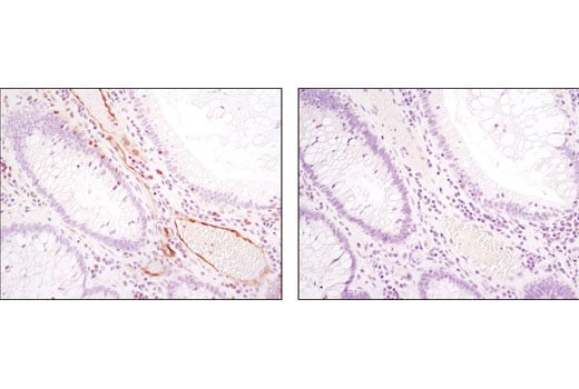
Immunohistochemical analysis of paraffin-embedded human colon (normal adjacent to tumor) using Phospho-PLCγ1 (Ser1248) (D25A9) Rabbit mAb in the presence of control peptide (left) or antigen-specific peptide (right).
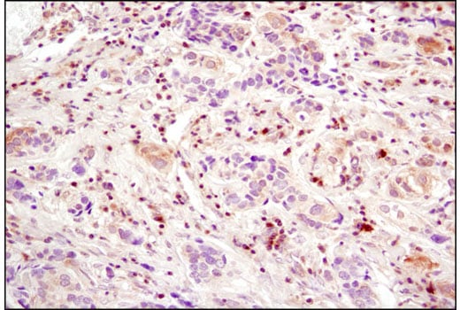
Immunohistochemical analysis of paraffin-embedded human breast carcinoma using Phospho-PLCγ1 (Ser1248) (D25A9) Rabbit mAb.
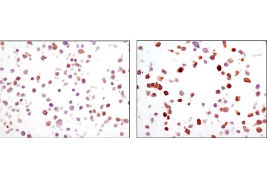
Immunohistochemical analysis of SignalSlide® Phospho-EGF Receptor IHC Controls #8102 [paraffin-embedded KYSE450 cell pellets untreated (left) or EGF-treated (right)] using Phospho-PLCγ1 (Ser1248) (D25A9) Rabbit mAb.
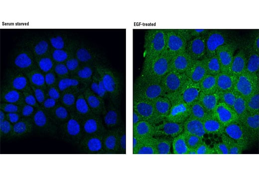
Confocal immunofluorescent analysis of A-431 cells, serum starved (left) or treated with hEGF #8916 (100 ng/mL for 15 min) using Phospho-PLCγ1 (Ser1248) (D25A9) Rabbit mAb (green). Blue pseudocolor = DRAQ5® #4084 (fluorescent DNA dye).
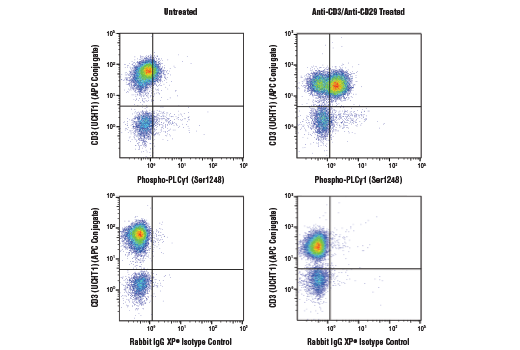
Flow cytometric analysis of human peripheral blood mononuclear cells, untreated (left column) or treated with cross-linked anti-CD3 plus anti-CD28 (10ug/ml each, 15 min; right column), using Phospho-PLCγ1 (Ser1248) (D25A9) Rabbit mAb (top row) or concentration-matched Rabbit (DA1E) mAb IgG XP® Isotype Control #3900 (bottom row), and co-stained with CD3 (UCHT1) Mouse mAb (APC Conjugate) #19881. Anti-rabbit IgG (H+L), F(ab')2 Fragment (Alexa Fluor® 488 Conjugate) #4412 was used as a secondary antibody.