


 全部商品分类


Phospho-Tau Family Antibody Sampler Kit

 下载产品说明书 下载SDS
 用小程序,查商品更便捷


 收藏
收藏
 对比
对比 咨询
咨询
The Phospho-Tau Family Antibody Sampler Kit provides an economical means of detecting the activation of Tau family members using phospho-specific and control antibodies. The kit includes enough antibody to perform two western blot experiments with each primary antibody.
参考图片
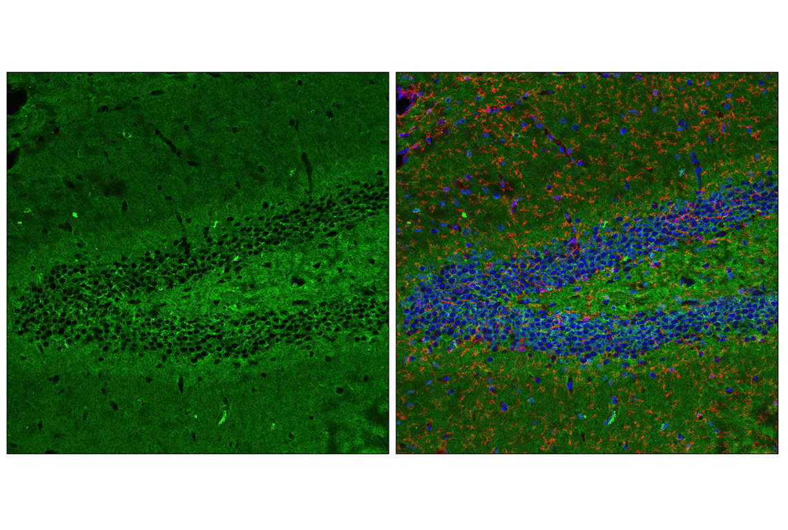
Confocal immunofluorescent analysis of fixed frozen mouse hippocampus using Tau (D1M9X) XP® Rabbit mAb (green), TMEM119 (E4B9S) Mouse mAb #98778 (red) and ProLong® Gold Antifade Reagent with DAPI #8961 (blue).
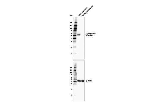
Western blot analysis of normal mouse brain and Tau KO (-/-) mouse brain with Phospho-Tau (Ser416) (D7U2P) Rabbit mAb (upper) and β-Actin (D6A8) Rabbit mAb #8457 (lower). Tau-KO mouse brain tissue was kindly provided by Dr. Dominic Walsh at Brigham and Women's Hospital and Harvard Medical School.
Western blot analysis of normal mouse brain and Tau KO (-/-) mouse brain with Phospho-Tau (Ser404) (D2Z4G) Rabbit mAb (upper) and β-Actin (D6A8) Rabbit mAb #8457 (lower). Tau-KO mouse brain tissue was kindly provided by Dr. Dominic Walsh at Brigham and Women's Hospital and Harvard Medical School.
Western blot analysis of normal mouse brain and Tau KO (-/-) mouse brain with Phospho-Tau (Ser404) (D2Z4G) Rabbit mAb (upper) and β-Actin (D6A8) Rabbit mAb #8457 (lower). Tau-KO mouse brain tissue was kindly provided by Dr. Dominic Walsh at Brigham and Women's Hospital and Harvard Medical School.
Western blot analysis of normal mouse brain and Tau KO (-/-) mouse brain with Phospho-Tau (Ser202) (D4H7E) Rabbit mAb (upper) and β-Actin (D6A8) Rabbit mAb #8457 (lower). Tau-KO mouse brain tissue was kindly provided by Dr. Dominic Walsh at Brigham and Women's Hospital and Harvard Medical School.
Western blot analysis of normal mouse brain and Tau KO (-/-) mouse brain with Tau (D1M9X) XP® Rabbit mAb (upper) and β-Actin (D6A8) Rabbit mAb #8457 (lower). Tau-KO mouse brain tissue was kindly provided by Dr. Dominic Walsh at Brigham and Women's Hospital and Harvard Medical School.
After the primary antibody is bound to the target protein, a complex with HRP-linked secondary antibody is formed. The LumiGLO® is added and emits light during enzyme catalyzed decomposition.
After the primary antibody is bound to the target protein, a complex with HRP-linked secondary antibody is formed. The LumiGLO* is added and emits light during enzyme catalyzed decomposition.
Western blot analysis of extracts from mouse brain and CAD cells, using Phospho-Tau (Ser396) (PHF13) Mouse mAb. The phospho-specificity of the antibody was verified by peptide blocking using no peptide, phospho-peptide or nonphospho-peptide.
Western blot analysis of mouse brain lysate, untreated (-) or λ phosphatase-treated (+), using Phospho-Tau (Ser416) (D7U2P) Rabbit mAb (upper) and β-Actin (D6A8) Rabbit mAb #8457 (lower).
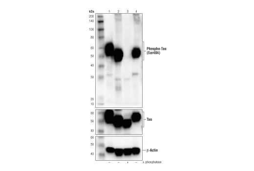
Western blot analysis of extracts from human cortex (lane 1), neonatal mouse brain, untreated (lane 2) or phosphatase-treated (lane 3), and fetal rat brain (lane 4), using Phospho-Tau (Ser404) (D2Z4G) Rabbit mAb (upper), Tau (Tau46) Mouse mAb #4019 (middle) and β-Actin (D6A8) Rabbit mAb #8457 (lower).
Western blot analysis of extracts from human cortex (lane 1), untreated neonatal mouse brain (lane 2), phosphatase-treated neonatal mouse brain (lane 3), and fetal rat brain (lane 4), using Phospho-Tau (Ser404) (D2Z4G) Rabbit mAb (upper), Tau (Tau46) Mouse mAb #4019 (middle), and β-Actin (D6A8) Rabbit mAb #8457 (lower).
Western blot analysis of extracts from mouse brain, untreated (-) or λ-phosphatase-treated (+), using Phospho-Tau (Ser202) (D4H7E) Rabbit mAb (upper) and β-Tubulin (D2N5G) Rabbit mAb #15115 (lower).
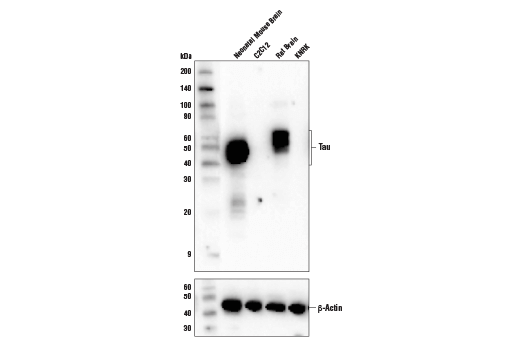
Western blot analysis of extracts from various cell lines and tissues using Tau (D1M9X) XP® Rabbit mAb (upper) and β-Actin (D6A8) Rabbit mAb #8457 (lower).
Western blot analysis of extracts from mouse and rat brain using Phospho-Tau (Ser416) (D7U2P) Rabbit mAb. The phospho-specificity of Phospho-Tau (Ser416) (D7U2P) Rabbit mAb was verified by peptide blocking using a phosphopeptide or non-phosphopeptide targeting residue Ser416.
Western blot analysis of extracts from MEF cells (lane 1) and mouse brain (lane 2) using Phospho-Tau (Ser404) (D2Z4G) Rabbit mAb (upper), Tau (Tau46) Mouse mAb #4019 (middle) and β-Actin (D6A8) Rabbit mAb #8457 (lower).
Immunohistochemical analysis of paraffin-embedded human Alzheimer's brain using Phospho-Tau (Ser404) (D2Z4G) Rabbit mAb.
Western blot analysis of extracts from neonatal and adult mouse brain using Phospho-Tau (Ser202) (D4H7E) Rabbit mAb. The phospho-specificity of the antibody was verified by blocking with a phospho- or nonphosphopeptide.
Immunohistochemical analysis of paraffin-embedded human Alzheimer's brain using Tau (D1M9X) XP® Rabbit mAb in the presence of control peptide (left) or antigen-specific peptide (right).
Immunohistochemical analysis of paraffin-embedded human breast carcinoma using Phospho-Tau (Ser416) (D7U2P) Rabbit mAb.
Confocal immunofluorescent analysis of Tg2576 mouse brain, untreated (left) or Lambda Protein Phosphatase-treated (right), using Phospho-Tau (Ser404) (D2Z4G) Rabbit mAb (red) and GFAP (GA5) Mouse mAb #3670 (green). Blue pseudocolor = DRAQ5® #4084 (fluorescent DNA dye).
Immunohistochemical analysis of paraffin-embedded mouse colon, untreated (left) or λ phosphatase-treated (right), using Phospho-Tau (Ser404) (D2Z4G) Rabbit mAb.
Immunohistochemical analysis of paraffin-embedded human normal appendix using Tau (D1M9X) XP® Rabbit mAb.
Immunohistochemical analysis of paraffin-embedded mouse brain, untreated (left) or λ phosphatase-treated (right), using Phospho-Tau (Ser416) (D7U2P) Rabbit mAb.
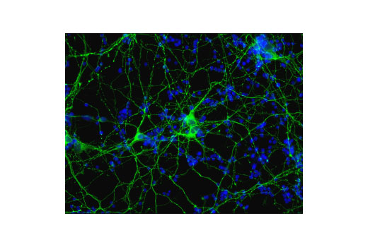
Confocal immunofluorescent analysis of mouse primary neurons using Phospho-Tau (Ser404) (D2Z4G) Rabbit mAb (green). Blue pseudocolor = Hoescht 33342 #4082 (fluorescent DNA dye).
Immunohistochemical analysis of paraffin-embedded T-47D cell pellets (left, positive) and MDA-MB-231 cell pellets (right, negative) using Phospho-Tau (Ser404) (D2Z4G) Rabbit mAb.
Immunohistochemical analysis of paraffin-embedded T-47D cell pellet (left, positive) or MDA-MB-231 cell pellet (right, negative) using Tau (D1M9X) XP® Rabbit mAb.
Immunohistochemical analysis of paraffin-embedded mouse brain using Phospho-Tau (Ser404) (D2Z4G) Rabbit mAb in the presence of non-phosphorylated tau peptide (left) and antigen-specific phospho-tau (Ser404) peptide (right).
Immunohistochemical analysis of paraffin-embedded mouse lung using Tau (D1M9X) XP® Rabbit mAb.
Confocal immunofluorescent analysis of fixed frozen mouse striatum using Tau (D1M9X) XP® Rabbit mAb (green), TMEM119 (E4B9S) Mouse mAb #98778 (red) and ProLong® Gold Antifade Reagent with DAPI #8961 (blue).
Confocal immunofluorescent analysis of T-47D (positive, left) or MDA-MB-231 (negative, right) cells using Tau (D1M9X) XP® Rabbit mAb (green). Blue pseudocolor = DRAQ5® #4084 (fluorescent DNA dye).
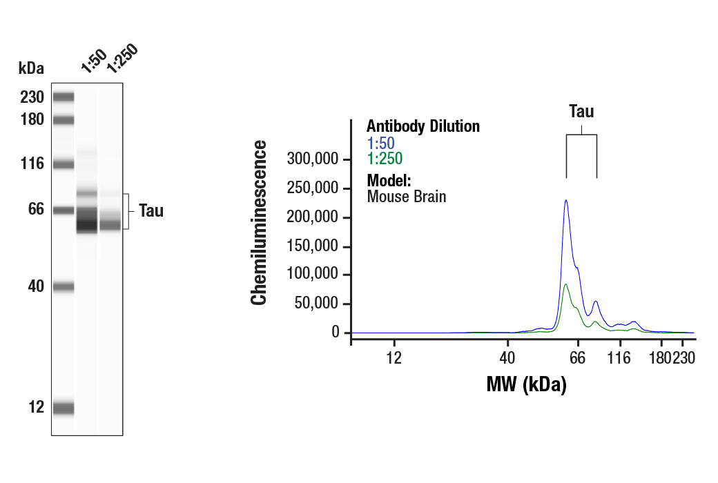
Simple Western™ analysis of lysates (0.1 mg/mL) from Mouse Brain Tissue Extracts using Tau (D1M9X) XP® Rabbit #46687. The virtual lane view (left) shows the target band (as indicated) and a band corresponding to Tau (as indicated) at 1:50 and 1:250 dilutions of primary antibody. The corresponding electropherogram view (right) plots chemiluminescence by molecular weight along the capillary at 1:50 (blue line) and 1:250 (green line) dilutions of primary antibody. This experiment was performed under reducing conditions on the Jess™ Simple Western instrument from ProteinSimple, a BioTechne brand, using the 12-230 kDa separation module.
Confocal immunofluorescent analysis of fixed frozen mouse cerebellum using Tau (D1M9X) XP® Rabbit mAb (green), TMEM119 (E4B9S) Mouse mAb #98778 (red) and ProLong® Gold Antifade Reagent with DAPI #8961 (blue).