


 全部商品分类


 下载产品说明书 下载SDS
 用小程序,查商品更便捷


 收藏
收藏
 对比
对比 咨询
咨询
The PI3 Kinase Sampler Kit provides an economical means of studying PI3 kinase subunits in cells. The kit contains enough primary and secondary antibodies to perform two Western blot experiments per primary antibody.
参考图片
After the primary antibody is bound to the target protein, a complex with HRP-linked secondary antibody is formed. The LumiGLO® is added and emits light during enzyme catalyzed decomposition.
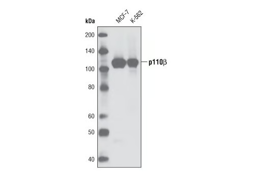
Western blot analysis of extracts from MCF-7 and K-562 cells using PI3 Kinase p110β (C33D4) Rabbit mAb.
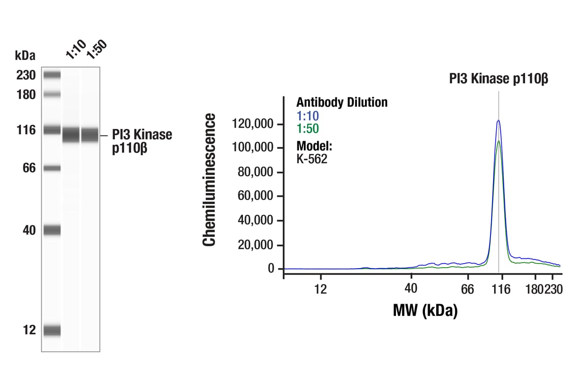
Simple WesternTM analysis of lysates (1 mg/ml) from K-562 cells lysates using PI3 Kinase p110β (C33D4) Rabbit mAb #3011. The virtual lane view (left) shows the target band (as indicated) at 1:10 and 1:50 dilutions of primary antibody. The corresponding electropherogram view (right) plots chemiluminescence by molecular weight along the capillary at 1:10 (blue line) and 1:50 (green line) dilutions of primary antibody. This experiment was performed under reducing conditions on the JessTM Simple Western instrument from ProteinSimple, a BioTechne brand, using the 12-230kDa.
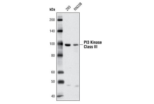
Western blot analysis of extracts from 293 and RN33B cells using PI3 Kinase Class III (D4E2) Rabbit mAb.
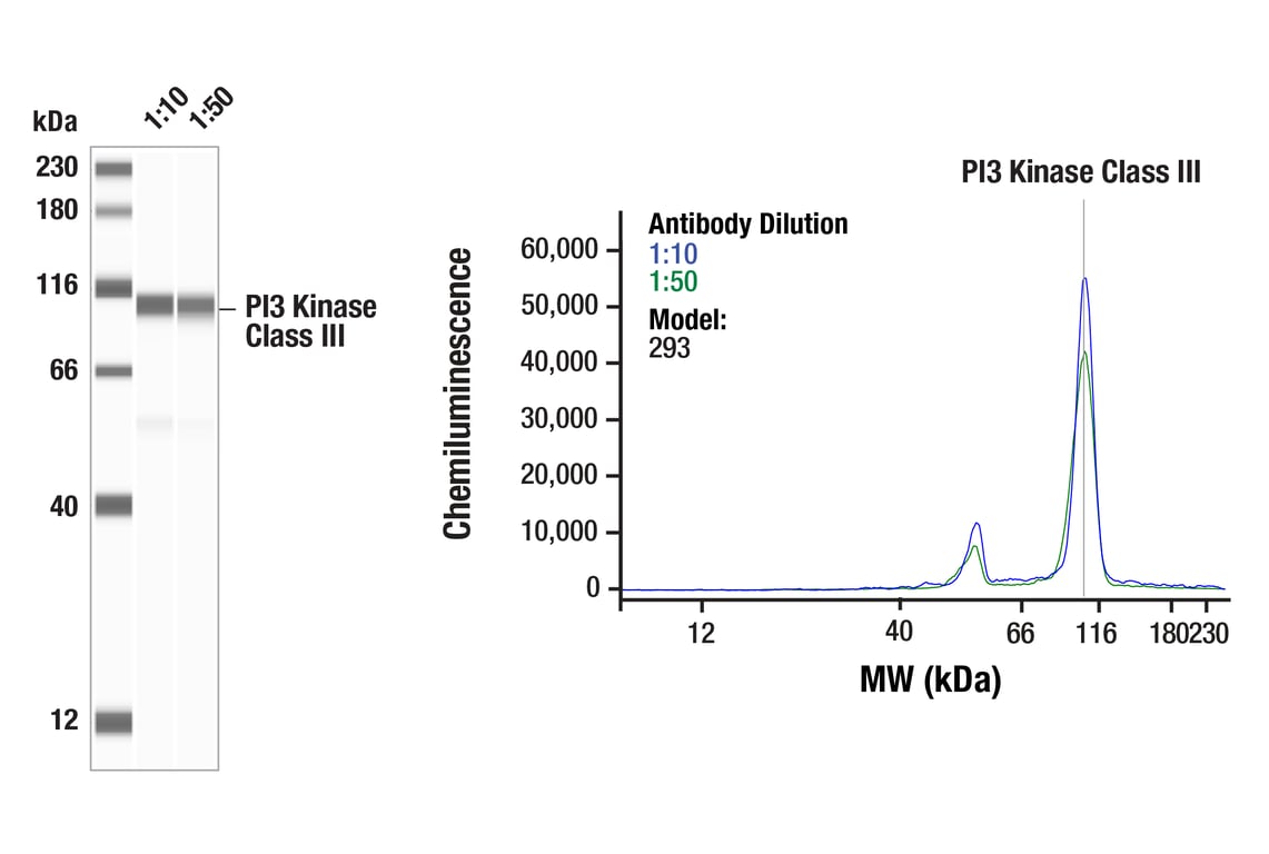
Simple Western™ analysis of lysates (1 mg/mL) from 293 cells using PI3 Kinase Class III (D4E2) Rabbit mAb #3358. The virtual lane view (left) shows the target band (as indicated) at 1:10 and 1:50 dilutions of primary antibody. The corresponding electropherogram view (right) plots chemiluminescence by molecular weight along the capillary at 1:10 (blue line) and 1:50 (green line) dilutions of primary antibody. This experiment was performed under reducing conditions on the Jess™ Simple Western instrument from ProteinSimple, a BioTechne brand, using the 12-230 kDa separation module.
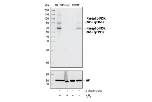
Western blot analysis of extracts from NIH/3T3-Src cells, untreated or treated with lambda phosphatase and from C2C12 cells, untreated or treated with H2O2, using Phospho-PI3 Kinase p85 (Tyr458)/p55 (Tyr199) Antibody.
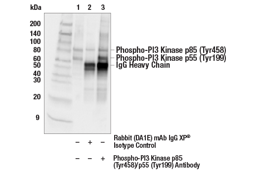
Immunoprecipitation of NIH/3T3-Src cell extracts. Lane 1 is 10% input, lane 2 is Rabbit (DA1E) mAb IgG XP® Isotype Control #3900, and lane 3 is Phospho-PI3 Kinase p85 (Tyr458)/p55 (Tyr199) Antibody. Western blot was performed using Phospho-PI3 Kinase p85 (Tyr458)/p55 (Tyr199) Antibody.
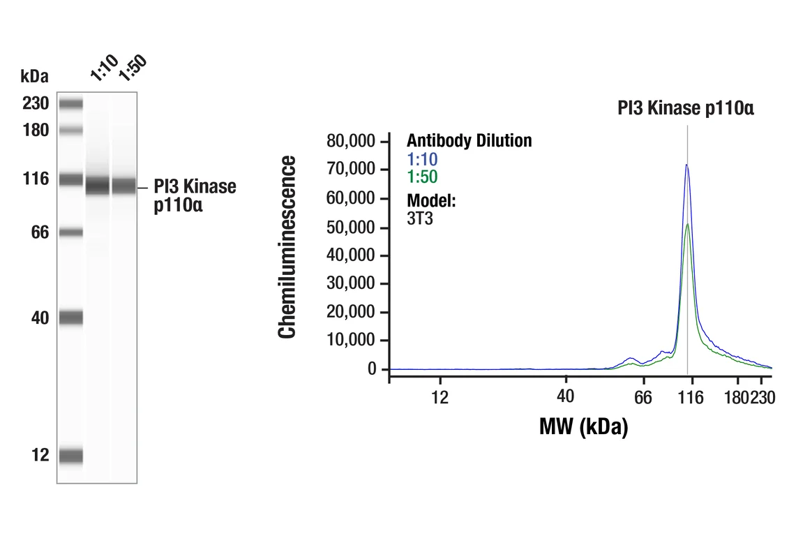
Simple Western™ analysis of lysates (1 mg/mL) from 3T3 cells using PI3 Kinase p110α (C73F8) Rabbit mAb #4249 . The virtual lane view (left) shows a single target band (as indicated) at 1:10 and 1:50 dilutions of primary antibody. The corresponding electropherogram view (right) plots chemiluminescence by molecular weight along the capillary at 1:10 (blue line) and 1:50 (green line) dilutions of primary antibody. This experiment was performed under reducing conditions on the Jess™ Simple Western instrument from ProteinSimple, a BioTechne brand, using the 12-230 kDa separation module.
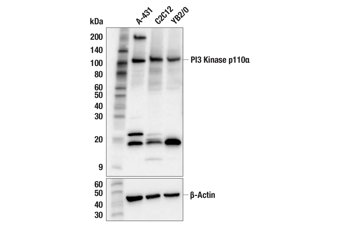
Western blot analysis of extracts from various cell lines using PI3 Kinase p110α (C73F8) Rabbit mAb (upper) or β-Actin (D6A8) Rabbit mAb #8457 (lower).
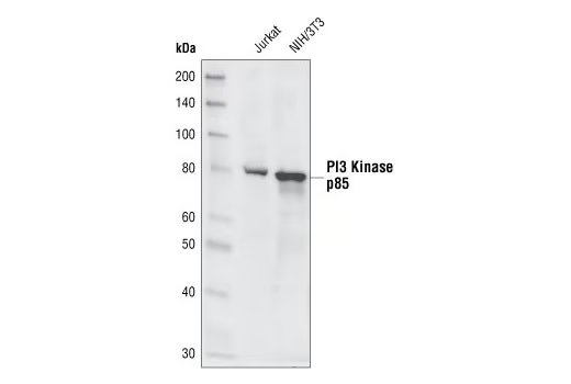
Western blot analysis of extracts from Jurkat and NIH/3T3 cells, using PI3 Kinase p85 (19H8) Rabbit mAb.
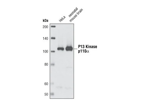
Western blot analysis of extracts from HeLa cells and neonatal mouse brain using PI3 Kinase p110α (C73F8) Rabbit mAb.
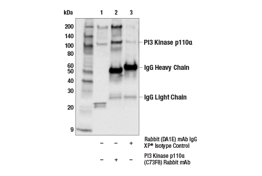
Immunoprecipitation of PI3 Kinase p110α protein from HeLa cell extracts. Lane 1 is 10% input, lane 2 is PI3 Kinase p110α (C73F8) Rabbit mAb, and lane 3 is Rabbit (DA1E) mAb IgG XP® Isotype Control #3900. Western blot analysis was performed using PI3 Kinase p110α (C73F8) Rabbit mAb. Anti-rabbit IgG, HRP-linked Antibody #7074 was used as a secondary antibody.
Simple Western™ analysis of lysates (1 mg/mL) from 3T3 cells using PI3 Kinase p85 (19H9) Rabbit mAb #4257. The virtual lane view (left) shows the target band (as indicated) at 1:10 and 1:50 dilutions of primary antibody. The corresponding electropherogram view (right) plots chemiluminescence by molecular weight along the capillary at 1:10 (blue line) and 1:50 (green line) dilutions of primary antibody. This experiment was performed under reducing conditions on the Jess™ Simple Western instrument from ProteinSimple, a BioTechne brand, using the 12-230 kDa separation module.
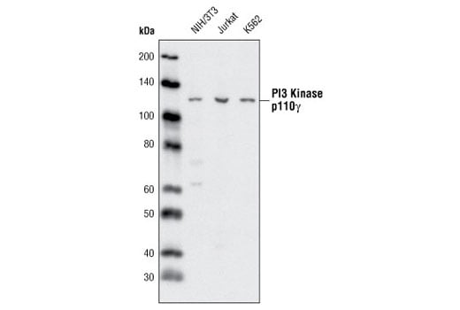
Western blot analysis of extracts from NIH/3T3, Jurkat and K562 cells using PI3 Kinase p110γ (D55D5) Rabbit mAb.