


 全部商品分类


PKM1 (D30G6) XP ® Rabbit mAb

 下载产品说明书 下载COA 下载SDS
 用小程序,查商品更便捷


 收藏
收藏
 对比
对比 咨询
咨询
Monoclonal antibody is produced by immunizing animals with a synthetic peptide corresponding to residues surrounding Asp407 of human PKM1 protein.
Product Usage Information
| Application | Dilution |
|---|---|
| Western Blotting | 1:1000 |
| Immunohistochemistry (Paraffin) | 1:300 - 1:1200 |
| Immunofluorescence (Frozen) | 1:200 - 1:400 |
| Immunofluorescence (Immunocytochemistry) | 1:200 - 1:400 |
| Flow Cytometry (Fixed/Permeabilized) | 1:400 - 1:1600 |
Specificity/Sensitivity
Species Reactivity:
Human, Mouse
Supplied in 10 mM sodium HEPES (pH 7.5), 150 mM NaCl, 100 µg/ml BSA, 50% glycerol and less than 0.02% sodium azide. Store at –20°C. Do not aliquot the antibody.
For a carrier free (BSA and azide free) version of this product see product #68774.
参考图片
Flow cytometric analysis of 293T cells mock transfected (blue) or transfected with hPKM1-Myc/DDK (green) using PKM1 (D30G6) XP® Rabbit mAb (solid lines) or concentration-matched Rabbit (DA1E) mAb IgG XP® Isotype Control #3900 (dashed lines).
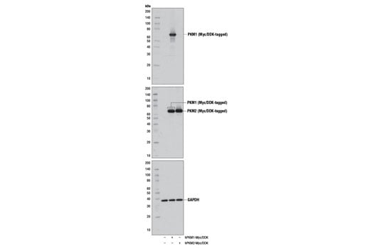
Western blot analysis of extracts from 293 cells, mock transfected (-) or transfected with a construct expressing Myc/DDK-tagged full-length human PKM1 (hPKM1-Myc/DDK; +) or Myc/DDK-tagged full-length human PKM2 (hPKM2-Myc/DDK; +), using PKM1 (D30G6) XP® Rabbit mAb (upper), DYKDDDDK (9A3) Mouse mAb #8146 (middle), or GAPDH (D16H11) XP® Rabbit mAb #5174 (lower).
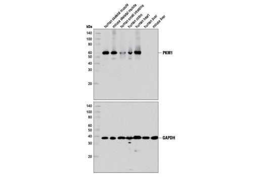
Western blot analysis of extracts from various mouse and human tissues using PKM1 (D30G6) XP® Rabbit mAb (upper) or GAPDH (D16H11) XP® Rabbit mAb #5174 (lower).
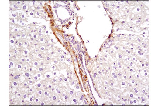
Immunohistochemical analysis of paraffin-embedded mouse liver using PKM1 (D30G6) XP® Rabbit mAb. Note the staining of vascular smooth muscle with no staining of hepatocytes.
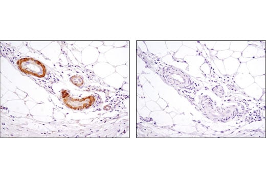
Immunohistochemical analysis of paraffin-embedded human colon using PKM1 (D30G6) XP® Rabbit mAb in the presence of control peptide (left) or antigen-specific peptide (right).
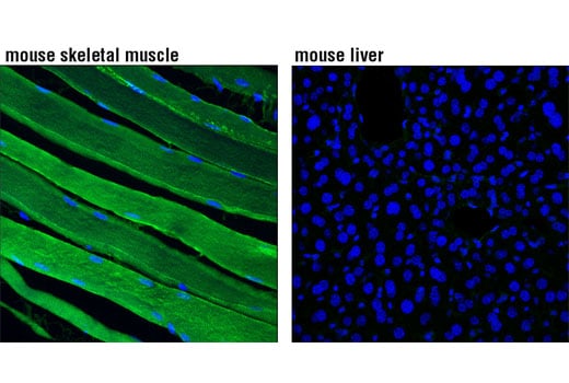
Confocal immunofluorescent analysis of mouse skeletal muscle (left) or liver (right) using PKM1 (D30G6) XP® Rabbit mAb (green). Blue pseudocolor = DRAQ5® #4084 (fluorescent DNA dye).
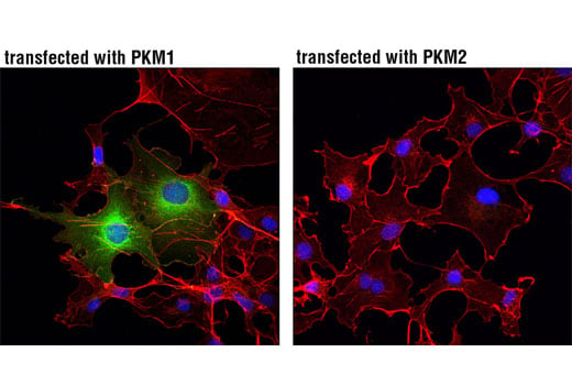
Confocal immunofluorescent analysis of COS-7 cells, transiently transfected with PKM1 (left) or PKM2 (right), using PKM1 (D30G6) XP® Rabbit mAb (green). Actin filaments were labeled with DyLight™ 554 Phalloidin #13054 (red). Blue pseudocolor = DRAQ5® #4084 (fluorescent DNA dye).