


 全部商品分类
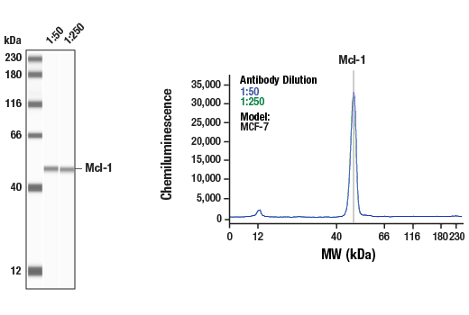


Pro-Survival Bcl-2 Family Antibody Sampler Kit II

 下载产品说明书 下载SDS
 用小程序,查商品更便捷


 收藏
收藏
 对比
对比 咨询
咨询
The Pro-Survival Bcl-2 Family Antibody Sampler Kit II provides an economical means to examine several members of the Bcl-2 family. The kit contains enough primary antibody to perform two western blot experiments.
参考图片
Simple Western™ analysis of lysates (1 mg/mL) from MCF-7 cells using Mcl-1 (D2W9E) Rabbit mAb #94296. The virtual lane view (left) shows the target band (as indicated) at 1:50 and 1:250 dilutions of primary antibody. The corresponding electropherogram view (right) plots chemiluminescence by molecular weight along the capillary at 1:50 (blue line) and 1:250 (green line) dilutions of primary antibody. This experiment was performed under reducing conditions on the Jess™ Simple Western instrument from ProteinSimple, a BioTechne brand, using the 12-230 kDa separation module.
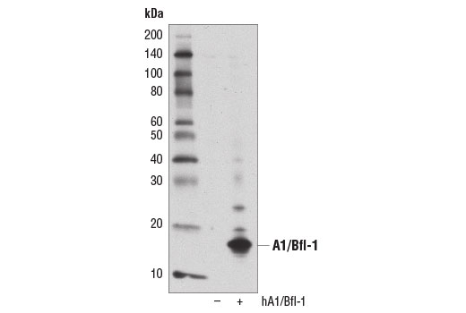
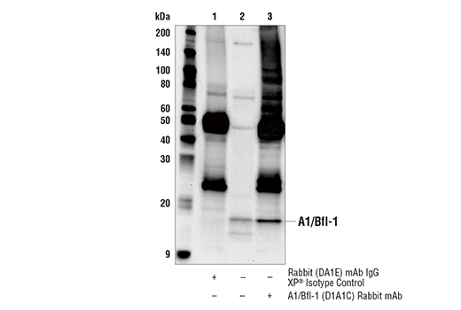
Western blot analysis of extracts from 293T cells, mock transfected (-) or transfected with a construct expressing full-length human A1/Bfl-1 protein (hA1/Bfl-1; +), using A1/Bfl-1 (D1A1C) Rabbit mAb.
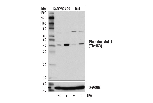
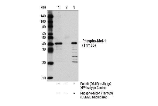
Western blot analysis of extracts from KARPAS-299 and Raji cell lines, untreated (-) or treated with TPA #4174 (200 nM, 30 min; +), using Phospho-Mcl-1 (Thr163) (D5M9D) Rabbit mAb (upper) and β-Actin (D6A8) Rabbit mAb #8457 (lower). KARPAS cell Line source: Dr Abraham Karpas at the University of Cambridge.
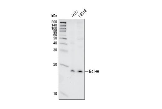
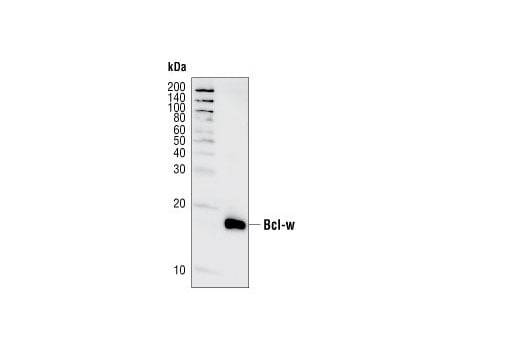
Western blot analysis of extracts from A673 and C2C12 cell lines, using Bcl-w (31H4) Rabbit mAb.
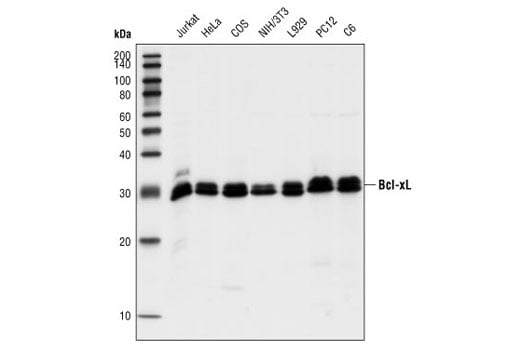
Western blot analysis of extracts from Jurkat and HeLa (human), COS (monkey), NIH/3T3 and L929 (mouse), and PC12 and C6 (rat) cells, using Bcl-xL (54H6) Rabbit mAb.
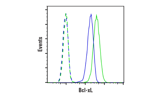
Flow cytometric analysis of THP-1 cells (blue, moderate expression) and K-562 cells (green, high expression) using Bcl-xL (54H6) Rabbit mAb (solid lines) or a concentration-matched Rabbit (DA1E) mAb IgG XP® Isotype Control #3900 (dashed lines). Anti-rabbit IgG (H+L), F(ab')2 Fragment (Alexa Fluor® 488 Conjugate) #4412 was used as a secondary antibody.
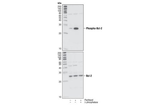
Western blot analysis of extracts from Jurkat cells, untreated or treated with paclitaxel (1 μM, overnight) and with or without λ phosphatase, using Phospho-Bcl-2 (Ser70) (5H2) Rabbit mAb (upper) or Bcl-2 #2876 (lower).
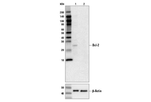
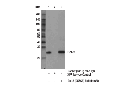
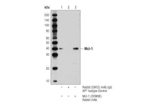
Western blot analysis of control HeLa cells (lane 1) or Bcl-2 knockout HeLa cells (lane 2) using Bcl-2 (D55G8) Rabbit mAb #4223 (upper), or β-actin (13E5) Rabbit mAb #4970 (lower). The absence of signal in the Bcl-2 knockout HeLa cells confirms the specificity of the antibody for Bcl-2.
After the primary antibody is bound to the target protein, a complex with HRP-linked secondary antibody is formed. The LumiGLO® is added and emits light during enzyme catalyzed decomposition.
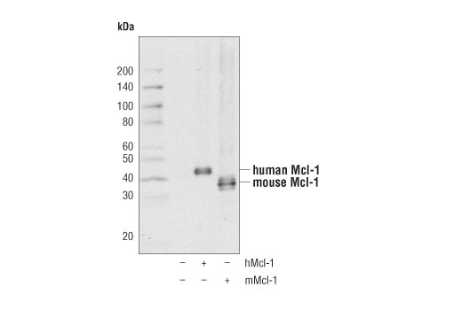
Western blot analysis of extracts from 293T cells, mock transfected or transfected with a construct expressing full-length human Mcl-1 (hMcl-1; +) or mouse Mcl-1 (mMcl-1; +), using Mcl-1 (D2W9E) Rabbit mAb.
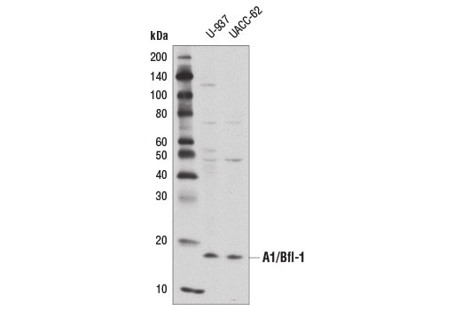
Western blot analysis of extracts from U-937 and UACC-62 cells using A1/Bfl-1 (D1A1C) Rabbit mAb.
Immunoprecipitation of phospho-Mcl-1 (Thr163) from KARPAS-299 cells treated with TPA #4174 (200 nM, 30 min) using Rabbit (DA1E) mAb IgG XP® Isotype Control #3900 (lane 2) or Phospho-Mcl-1 (Thr163) (D5M9D) Rabbit mAb (lane 3). Lane 1 represents 10% input. Western blot was perform using Phospho-Mcl-1 (Thr163) (D5M9D) Rabbit mAb. Mouse Anti-rabbit IgG (Conformation Specific) (L27A9) mAb #3678 was used as a secondary antibody to avoid cross reactivity with IgG heavy and light chains. KARPAS cell Line source: Dr Abraham Karpas at the University of Cambridge.
Western blot analysis of recombinant Bcl-w (amino acids 1-172) using Bcl-w (31H4) Rabbit mAb.
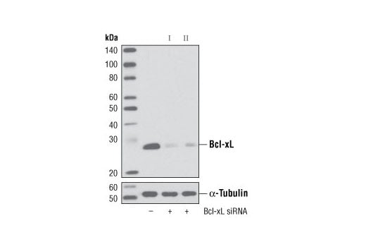
Western blot analysis of extracts from HeLa cells, transfected with 100 nM SignalSilence® Control siRNA (Unconjugated) #6568 (-), SignalSilence® BcL-xL siRNA I (+) or SignalSilence® Bcl-xL siRNA II #6363 (+), using Bcl-xL (54H6) Rabbit mAb #2764 (upper) or α-Tubulin (11H10) Rabbit mAb #2125 (lower). The Bcl-xL (54H6) Rabbit mAb confirms silencing of Bcl-xL expression, while the α-Tubulin (11H10) Rabbit mAb is used as a loading control.
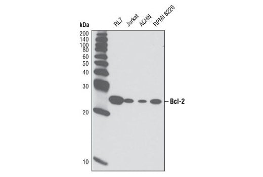
Western blot analysis of extracts from various cell lines using Bcl-2 (D55G8) Rabbit mAb.
Immunoprecipitation of A1/Bfl-1 from Mutz-3 cell extracts. Lane 1 is Rabbit (DA1E) mAb IgG XP® Isotype Control #3900, lane 2 is 10% input, and lane 3 is A1/Bfl-1 (D1A1C) Rabbit mAb #14093. Western blot analysis was performed using A1/Bfl-1 (D1A1C) Rabbit mAb #14093. Anti-rabbit IgG, HRP-linked Antibody #7074 was used as the secondary antibody.
Immunoprecipitation of Bcl-2 from ACHN extracts. Lane 1 is 10% input, lane 2 is Rabbit (DA1E) mAb IgG XP® Isotype Control #3900, and lane 3 is Bcl-2 (D55G8) Rabbit mAb. Western blot analysis was performed using Bcl-2 (124) Mouse mAb #15071. Anti-mouse IgG, HRP-linked Antibody #7076 was used as a secondary antibody.
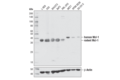
Western blot analysis of extracts from various cell lines using Mcl-1 (D2W9E) Rabbit mAb (upper) or β-Actin (D6A8) Rabbit mAb #8457 (lower).
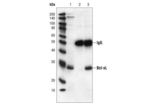
Immunoprecipitation of Bcl-xL from Jurkat cell extracts, using Bcl-xL (54H6) Rabbit mAb. Lane 1 is the lysate control, lane 2 is antibody alone and lane 3 is antibody plus lysate.
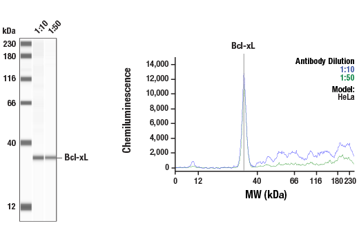
Simple Western™ analysis of lysates (1.0 mg/mL) from HeLa cells using Bcl-xL (54H6) Rabbit mAb #2764. The virtual lane view (left) shows a single target band (as indicated) at 1:10 and 1:50 dilutions of primary antibody. The corresponding electropherogram view (right) plots chemiluminescence by molecular weight along the capillary at 1:10 (blue line) and 1:50 (green line) dilutions of primary antibody. This experiment was performed under reducing conditions on the Jess™ Simple Western instrument from ProteinSimple, a BioTechne brand, using the 12-230 kDa separation module.
Immunoprecipitation of Mcl-1 from MCF7 cell extracts. Lane 1 is 10% input, lane 2 is Rabbit (DA1E) mAb IgG XP® Isotype Control #3900, and lane 3 is Mcl-1 (D2W9E) Rabbit mAb. Western blot analysis was performed using Mcl-1 (D2W9E) Rabbit mAb. A conformation-specific secondary antibody was used to avoid cross-reactivity with IgG.
Immunohistochemical analysis of paraffin-embedded human lung carcinoma, using Bcl-xL (54H6) Rabbit mAb.
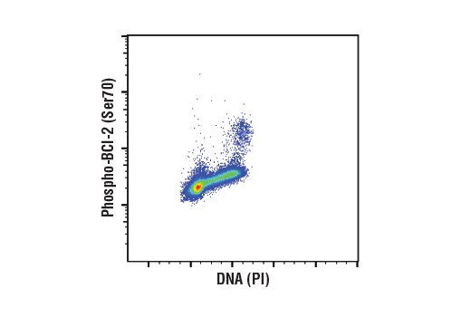
Flow cytometric analysis of Jurkat cells using Phospho-Bcl-2 (Ser70) (5H2) Rabbit mAb and Propidium Iodide (PI)/RNase Staining Solution #4087 to measure DNA content. Anti-rabbit IgG (H+L), F(ab')2 Fragment (Alexa Fluor® 488 Conjugate) #4412 was used as a secondary antibody.
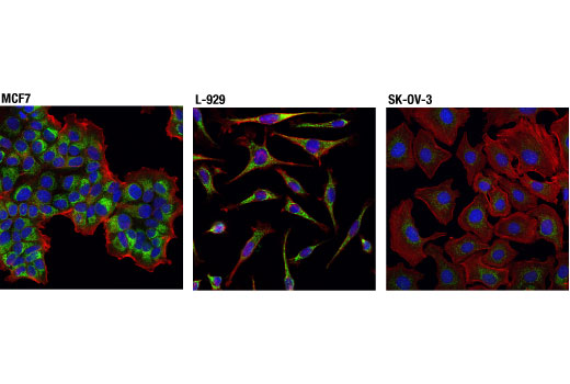
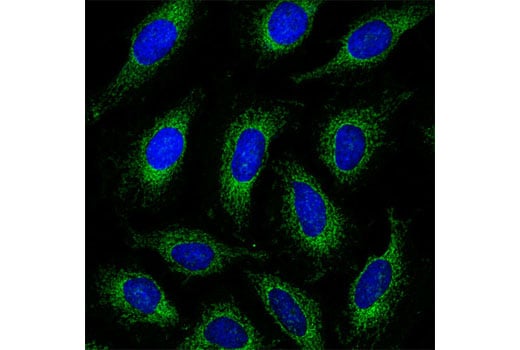
Confocal immunofluorescent analysis of MCF7 (left), L-929 (center), and SK-OV-3 (right) cells using Mcl-1 (D2W9E) Rabbit mAb (green). Actin filaments were labeled with DyLight™ 554 Phalloidin #13054 (red). Blue pseudocolor = DRAQ5® #4084 (fluorescent DNA dye).
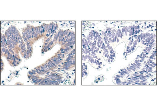
Immunohistochemical analysis of paraffin-embedded human colon carcinoma, using Bcl-xL (54H6) Rabbit mAb in the presence of control peptide (left) or Bcl-xL Blocking Peptide #1225 (right).
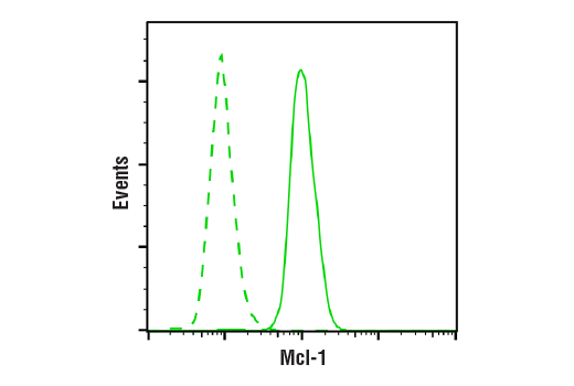
Flow cytometric analysis of L-929 cells using Mcl-1 (D2W9E) Rabbit mAb (solid line) compared to concentration matched Rabbit (DA1E) mAb IgG XP® Isotype Control #3900 (dashed line). Anti-rabbit IgG (H+L), F(ab')2 Fragment (Alexa Fluor® 488 Conjugate) #4412 was used as a secondary antibody.
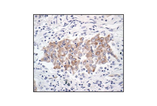
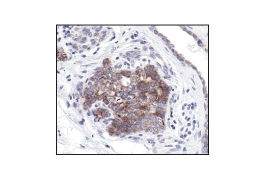
Immunohistochemical analysis of paraffin-embedded human prostate carcinoma, showing cytoplasmic localization, using Bcl-xL (54H6) Rabbit mAb.
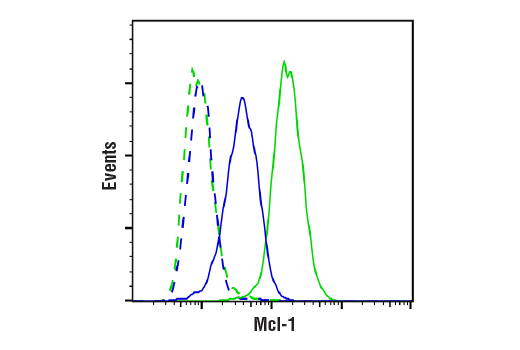
Flow cytometric analysis of SK-OV-3 cells (low expression; blue) and MCF7 cells (high expression; green) using Mcl-1 (D2W9E) Rabbit mAb (solid lines) or a concentration-matched Rabbit (DA1E) mAb IgG XP® Isotype Control #3900 (dashed lines). Anti-rabbit IgG (H+L), F(ab')2 Fragment (Alexa Fluor® 488 Conjugate) #4412 was used as a secondary antibody.
Confocal immunofluorescent analysis of HeLa cells using Bcl-xL (54H6) Rabbit mAb (green). Blue pseudocolor = DRAQ5® #4084 (fluorescent DNA dye).