


 下载产品说明书
 用小程序,查商品更便捷


 收藏
收藏
 对比
对比 咨询
咨询
Specificity/Sensitivity
参考图片
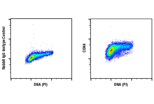
Flow cytometric analysis of Jurkat cells using CDK4 (D9G3E) Rabbit mAb and Propidium Iodid (PI)/RNase Staining Solution #4087 to measure DNA content. Anti-rabbit IgG (H+L), F(ab')2 Fragment (Alexa Fluor® 488 Conjugate) #4412 was used as a secondary antibody.
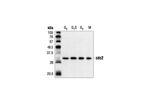
Western blot analysis of extracts from HeLa cells synchronized at various stages of the cell cycle, using cdc2 (POH1) Mouse mAb.
After the primary antibody is bound to the target protein, a complex with HRP-linked secondary antibody is formed. The LumiGLO* is added and emits light during enzyme catalyzed decomposition.
After the primary antibody is bound to the target protein, a complex with HRP-linked secondary antibody is formed. The LumiGLO* is added and emits light during enzyme catalyzed decomposition.
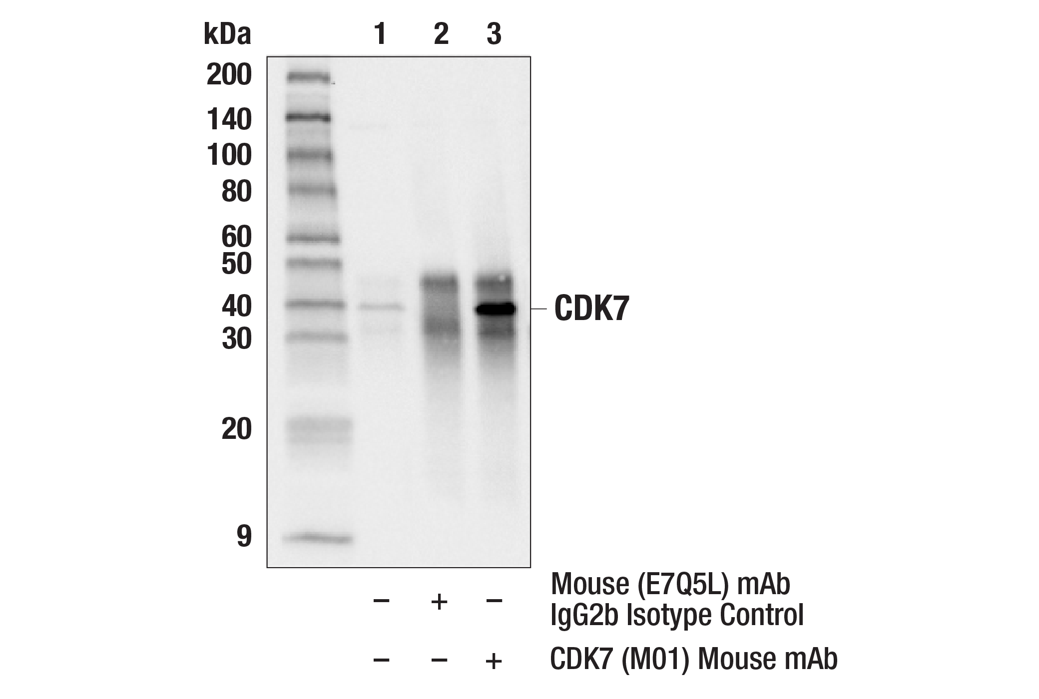
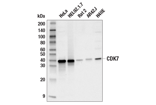
Western blot analysis of extracts from SK-N-MC and C6 cells, using CDK7 (MO1) Mouse mAb.
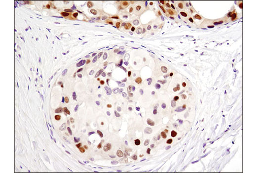
Immunohistochemical analysis of paraffin-embedded human breast carcinoma, showing nuclear localization, using CDK7 (MO1) Mouse mAb.
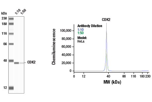
Western blot analysis of extracts from HeLa, NIH/3T3, C6 and COS cells, using CDK2 (78B2) Rabbit mAb.
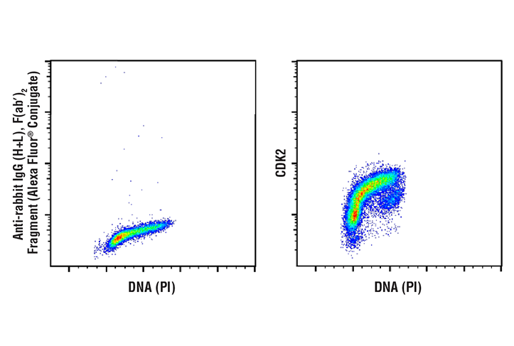
Flow cytometric analysis of Jurkat cells, using CDK2 (78B2) Rabbit mAb versus propidium iodide (DNA content).
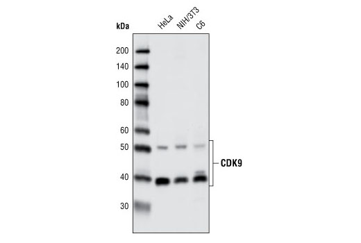
Western blot analysis of extracts from various cell types using CDK9 (C12F7) Rabbit mAb.
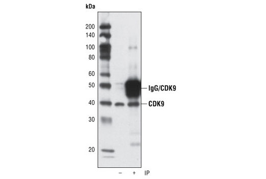
Immunoprecipitation of CDK9 from HeLa cells using CDK9 (C12F7) Rabbit mAb. Western blot detection was performed using the same antibody. Lane 1 is 5% input.
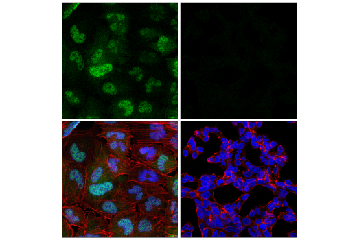
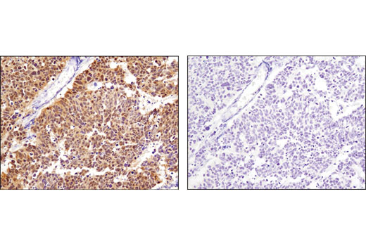
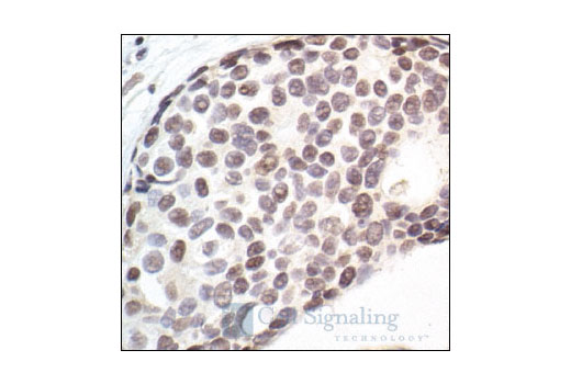
Immunohistochemical analysis of paraffin-embedded human breast carcinoma using CDK9 (C12F7) Rabbit mAb in the presence of control peptide (left) or antigen specific peptide (right).
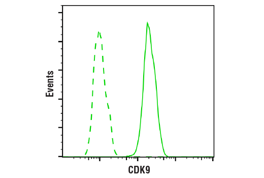
Flow cytometric analysis of Jurkat cells using CDK9 (C12F7) Rabbit mAb (blue) compared to a nonspecific negative control antibody (red).
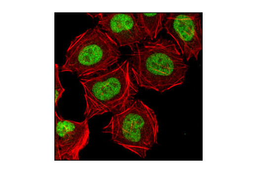
Confocal immunofluorescent analysis of HeLa cells using CDK9 (C12F7) Rabbit mAb (green). Actin filaments have been labeled with DY-555 phalloidin (red).
Immunohistochemical analysis of paraffin-embedded K7M2 mouse syngeneic tumor using CDK9 (C12F7) Rabbit mAb.
Immunohistochemical analysis of frozen SKOV-3 xenograft using CDK9 (C12F7) Rabbit mAb.
Western blot analysis of extracts from SK-N-MC and C6 cells using CDK7 (MO1) Mouse mAb #2916.Western blot方法检测SK-N-MC和 C6细胞提取物,使用的抗体为 CDK7 (MO1) Mouse mAb #2916。
Western blot analysis of extracts from various cell types using CDK2 (78B2) Rabbit mAb #2546.Western blot 检测不同细胞提取物,使用的抗体为CDK2 (78B2) Rabbit mAb #2546。
Western blot analysis of extracts from HeLa, NIH/3T3 and C6 cells using CDK9 (C12F7) Rabbit mAb #2316.Western blot方法检测HeLa, NIH/3T3 和C6 细胞提取物,使用的抗体为CDK9 (C12F7) Rabbit mAb #2316。
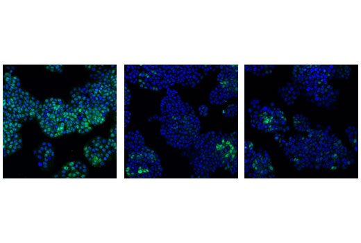
Confocal immunofluorescent analysis of HT-29 cells, transfected with SignalSilence® Control siRNA (Unconjugated) #6568 (left), SignalSilence® cdc2 siRNA I #3500 (center) or SignalSilence® cdc2 siRNA II #3600 (right), using cdc2 (POH1) Mouse mAb (green). Blue pseudocolor = DRAQ5® #4084 (fluorescent DNA dye).
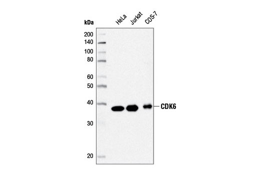
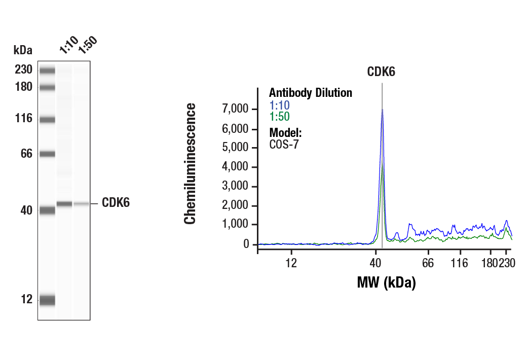
Western blot analysis of extracts from HeLa, Jurkat, and COS-7 cells using CDK6 (D4S8S) Rabbit mAb #13331.
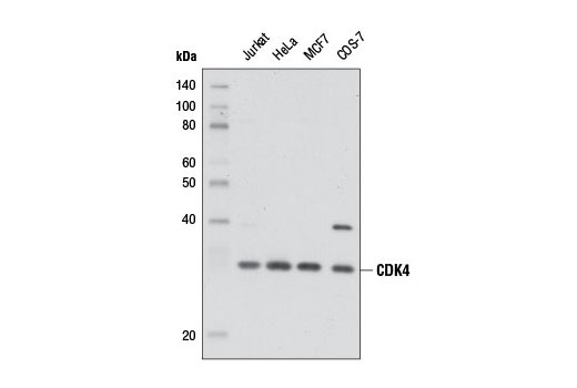
Western blot analysis of extracts from various cell lines using CDK4 (D9G3E) Rabbit mAb #12790.
Western blot analysis of extracts from HeLa cells synchronized at various stages of the cell cycle, using cdc2 (POH1) Mouse mAb #9116.
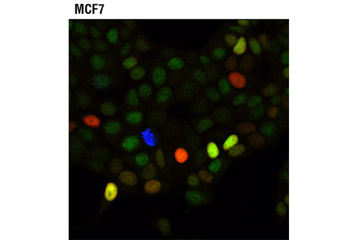
Confocal immunofluorescent analysis of MCF7 cells using CDK4 (D9G3E) Rabbit mAb (green), p21 Waf1/Cip1 (12D1) Rabbit mAb (Alexa Fluor® 555 Conjugate) #8493 (red), and Phospho-Histone H3 (Ser10) (D2C8) XP® Rabbit mAb (Alexa Fluor® 647 Conjugate) #3458 (blue pseudocolor).
Western blot analysis of extracts from various cell lines using CDK4 (D9G3E) Rabbit mAb.
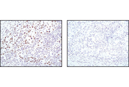
Immunohistochemical analysis of paraffin-embedded human lung carcinoma using CDK4 (D9G3E) Rabbit mAb in the presence of control peptide (left) or antigen-specific peptide (right).
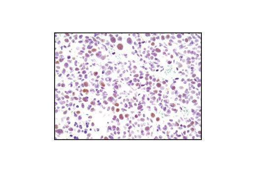
Immunohistochemical analysis of paraffin-embedded human breast carcinoma using CDK4 (D9G3E) Rabbit mAb.
Western blot analysis of extracts from HeLa, Jurkat, and COS-7 cells using CDK6 (D4S8S) Rabbit mAb.
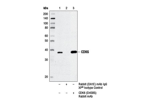
Immunoprecipitation of CDK6 from HeLa cell extracts using Rabbit (DA1E) mAb IgG XP® Isotype Control #3900 (lane 2) or CDK6 (D4S8S) Rabbit mAb (lane 3). Lane 1 is 10% input. Western blot was performed using CDK6 (DCS83) Mouse mAb #3136.

 危险品化学品经营许可证(不带存储) 许可证编号:沪(杨)应急管危经许[2022]202944(QY)
  营业执照(三证合一)