


 全部商品分类
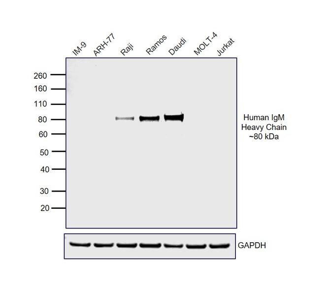


GOXHU IGM HRP

 下载产品说明书 下载COA 下载SDS
 用小程序,查商品更便捷


 收藏
收藏
 对比
对比 咨询
咨询免疫印迹 (WB)
免疫组化 (IHC)
免疫细胞化学 (ICC/IF)
种属反应
宿主/亚型
分类
类型
偶联物
形式
浓度
纯化类型
保存液
内含物
保存条件
运输条件
RRID
靶标
抗体形式
产品详细信息
Concentration may vary slightly from lot-to-lot, see lot-specific datasheet for exact concentration.
This antibody has been successfully used in Western blot, and ICC applications.
Antibody Specificity: This antibody reacts with the Fc5µ portion of the human IgM heavy chain, based on electrophoresis. No antibody was detected against normal human IgG or IgA, or against non-immunoglobulin serum proteins. However, this antibody may cross-react with IgM from other species.
Restoration and Storage: Store product at 4°C until opened. Restore with 2.0 mL distilled water (0.8 mg/mL after restoration). Centrifuge product if it is not completely clear after standing for 1-2 hours at room temperature. To judge clarity, draw product into a pasteur pipette. Product may be stored for several weeks at 4°C as an undiluted liquid. After dilution, do not use for more than one day.
To extend the shelf-life of this product, add an equal volume of glycerol to make a final concentration of approximately 50% glycerol and store at -20°C.
Country of Origin: USA
靶标信息
Anti-Human secondary antibodies are affinity-purified antibodies with well-characterized specificity for human immunoglobulins and are useful in the detection, sorting or purification of its specified target. Secondary antibodies offer increased versatility enabling users to use many detection systems (e.g. HRP, AP, fluorescence). They can also provide greater sensitivity through signal amplification as multiple secondary antibodies can bind to a single primary antibody. Most commonly, secondary antibodies are generated by immunizing the host animal with a pooled population of immunoglobulins from the target species and can be further purified and modified (i.e. immunoaffinity chromatography, antibody fragmentation, label conjugation, etc.) to generate highly specific reagents.
仅用于科研。不用于诊断过程。未经明确授权不得转售。
参考图片
Western blot was performed using Goat anti-Human IgM Secondary Antibody, HRP (Product # 31415) and an ~80 kDa band corresponding to Human IgM Heavy Chain was observed in Raji, Ramos and Daudi but not in IM-9, ARH-77, MOLT-4 and Jurkat. Whole cell extracts (30 µg) of IM-9 (Lane 1), ARH-77 (Lane 2), Raji (Lane 3), Ramos (Lane 4), Daudi (Lane 5), MOLT-4 (Lane 6) and Jurkat (Lane 7) were electrophoresed using NuPAGE™ 4-12% Bis-Tris Protein Gel (Product # NP0322BOX). Resolved proteins were then transferred onto a nitrocellulose membrane (Product # IB23001) by iBlot® 2 Dry Blotting System (Product # IB21001). The blot was probed with Goat anti-Human IgM Secondary Antibody, HRP (Product # 31415) (1:5000 dilution) and detected using the iBright FL1500 (Product # A44115). Chemiluminescent detection was performed using Novex® ECL Chemiluminescent Substrate Reagent Kit (Product # WP20005). Raji, Ramos and Daudi are known to express IgM whereas IM-9 and ARH-77 express IgG and are negative for IgM. MOLT-4 and Jurkat, being T-cell lines, do not express immunoglobulins. (DOI:10.1002/eji.1830100305; 10.3791/3573; 10.1016/0022-1759(94)00286-6; PMID: 566614).
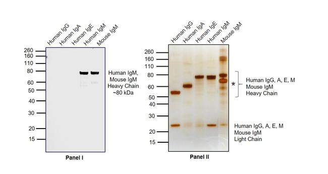
Western blot was performed using Goat anti-Human IgM Secondary Antibody, HRP (Product # 31415) and a ~80 kDa band corresponding to Heavy Chain were observed in Human IgM and Mouse IgM but not in Human IgG, IgA and IgE (Panel I). Purified protein (100 ng) of Human IgG (Lane 1), IgA (Lane 2), IgE (Lane 3), IgM (Lane 4) and Mouse IgM (Lane 5) were electrophoresed using NuPAGE™ 4-12% Bis-Tris Protein Gel (Product # NP0322BOX). Resolved proteins were then transferred onto a nitrocellulose membrane (Product # IB23001) by iBlot® 2 Dry Blotting System (Product # IB21001). The blot was probed with Product # 31415 (1:5000 dilution) and detected by chemiluminescence using Novex® ECL Chemiluminescent Substrate Reagent Kit (Product # WP20005) using the iBright FL1500 (Product # A44115). Silver staining was performed to establish equivalent loading of purified proteins using the Pierce™ Silver Stain Kit (Product # 24612) (Panel II). A band corresponding to BSA (*) which is part of the Mouse IgM formulation can be seen at ~60 kDa.