


 全部商品分类
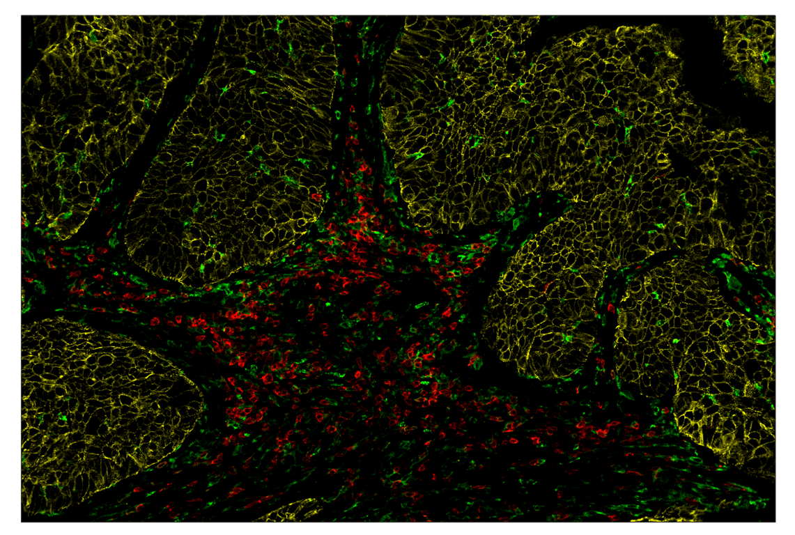


 下载产品说明书 下载SDS
 用小程序,查商品更便捷


 收藏
收藏
 对比
对比 咨询
咨询Monoclonal antibody is produced by immunizing animals with a synthetic peptide corresponding to residues surrounding Pro780 of human E-cadherin protein.

SignalStar multiplex immunohistochemistry (IHC) is an advanced technology for labeling multiple proteins simultaneously in tissue samples using specific primary antibodies and fluorescent detection reagents. This technology offers accuracy and reliability in visualizing and analyzing protein expression while maintaining spatial context and tissue architecture.
SignalStar Oligo-Antibody Pairs are compatible with the SignalStar Multiplex IHC Buffer Kits for use in fluorescent multiplex imaging experiments. This product includes the oligo-conjugated antibodies and complementary oligos required for labeling your target protein on up to 10 slides. SignalStar Multiplex IHC Buffer Kits are required to amplify and image the target signal. Multiple oligo-antibody pairs can be conveniently combined into a multiplex panel using the SignalStar Multiplex IHC Panel Builder. SignalStar Multiplex IHC Kits & Reagents are not compatible with all of Cell Signaling Technology® products and protocols that are recommended for use in immunohistochemical assays.
Product Usage Information
| Application | Dilution |
|---|---|
| SignalStar™ Leica Bond | 1:50 - 1:200 |
| SignalStar™ Manual | 1:50 - 1:200 |
Specificity/Sensitivity
Species Reactivity:
Human, Mouse
SignalStar conjugates are supplied in PBS (pH 7.2), less than 0.1% sodium azide, 2 mM EDTA, 0.05% Triton X-100, 2 mg/mL BSA, and 50% glycerol. Complementary oligos are supplied in nuclease-free water. Store at -20°C. Do not aliquot the antibody. All components in this kit are stable for at least 12 months when stored at the recommended temperature.
参考图片
SignalStar™ multiplex immunohistochemical analysis of paraffin-embedded human squamous cell lung carcinoma using MHC Class II (LGII-612.14) & CO-0105-488 SignalStar™ Oligo-Antibody Pair #55436 (green), E-Cadherin (24E10) & CO-0103-594 SignalStar™ Oligo-Antibody Pair #65400 (yellow), and CD28 (D2Z4E) & CO-0081-647 SignalStar™ Oligo-Antibody Pair #96977 (red). All fluorophores have been assigned a pseudocolor, as indicated.
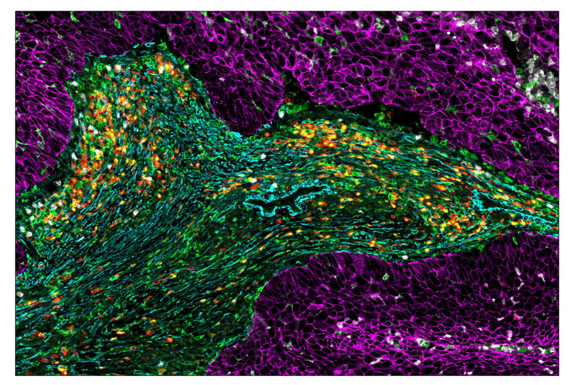
SignalStar™ multiplex immunohistochemical analysis of paraffin-embedded human squamous cell lung carcinoma using MHC Class II (LGII-612.14) & CO-0105-488 SignalStar™ Oligo-Antibody Pair #55436 (green), E-Cadherin (24E10) & CO-0103-594 SignalStar™ Oligo-Antibody Pair #65400 (magenta), CD28 (D2Z4E) & CO-0081-647 SignalStar™ Oligo-Antibody Pair #96977 (red), α-Smooth Muscle Actin (D4K9N) & CO-0024-488 SignalStar™ Oligo-Antibody Pair #76133 (green), TCF1/TCF7 (C63D9) & CO-0006-594 SignalStar™ Oligo-Antibody Pair #34706 (yellow), and Arginase-1 (D4E3M) & CO-0075-647 SignalStar™ Oligo-Antibody Pair #44900 (white). All fluorophores have been assigned a pseudocolor, as indicated. Staining was performed on the BOND RX autostainer by Leica Biosystems.
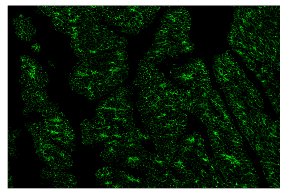
SignalStar™ multiplex immunohistochemical analysis of paraffin-embedded human colorectal adenocarcinoma using E-Cadherin (24E10) & CO-0103-488 SignalStar™ Oligo-Antibody Pair #48503 (green). All fluorophores have been assigned a pseudocolor, as indicated. Staining was performed on the BOND RX autostainer by Leica Biosystems.
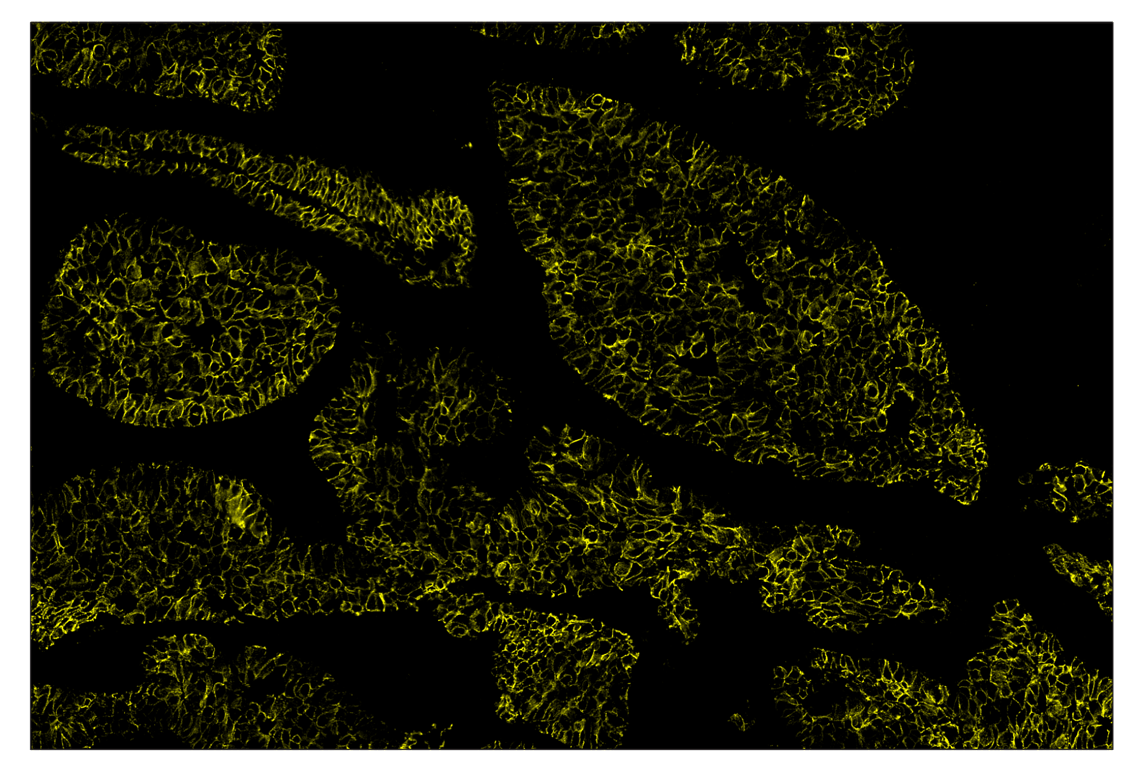
SignalStar™ multiplex immunohistochemical analysis of paraffin-embedded human lung adenocarcinoma using E-Cadherin (24E10) & CO-0103-594 SignalStar™ Oligo-Antibody Pair #65400 (yellow). All fluorophores have been assigned a pseudocolor, as indicated. Staining was performed on the BOND RX autostainer by Leica Biosystems.
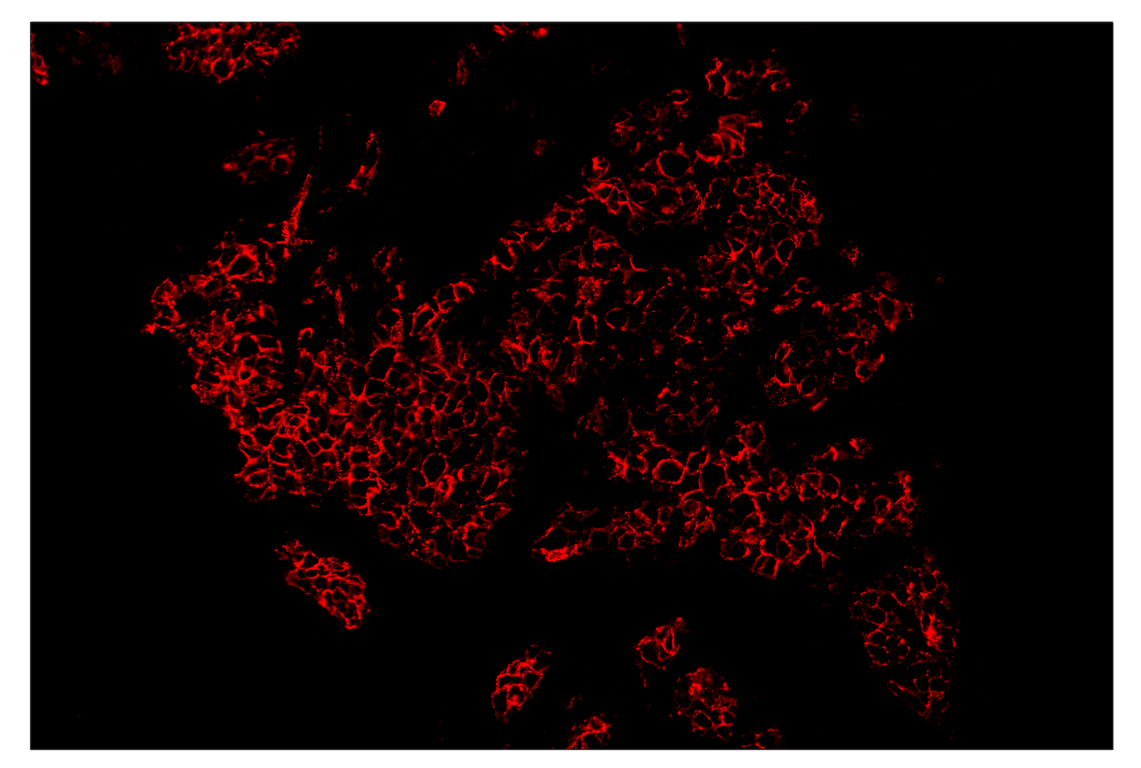
SignalStar™ multiplex immunohistochemical analysis of paraffin-embedded human non-small cell lung carcinoma using E-Cadherin (24E10) & CO-0103-647 SignalStar™ Oligo-Antibody Pair #31774 (red). All fluorophores have been assigned a pseudocolor, as indicated. Staining was performed on the BOND RX autostainer by Leica Biosystems.
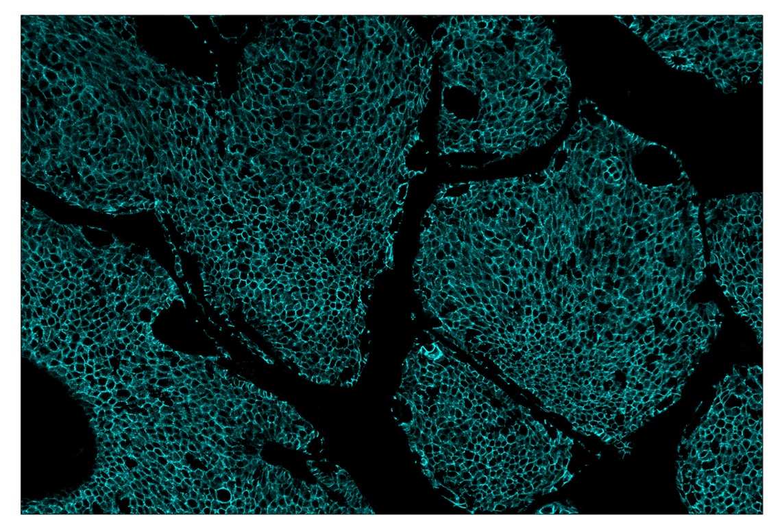
SignalStar™ multiplex immunohistochemical analysis of paraffin-embedded human squamous cell lung carcinoma using E-Cadherin (24E10) & CO-0103-750 SignalStar™ Oligo-Antibody Pair #65506 (cyan). All fluorophores have been assigned a pseudocolor, as indicated. Staining was performed on the BOND RX autostainer by Leica Biosystems.
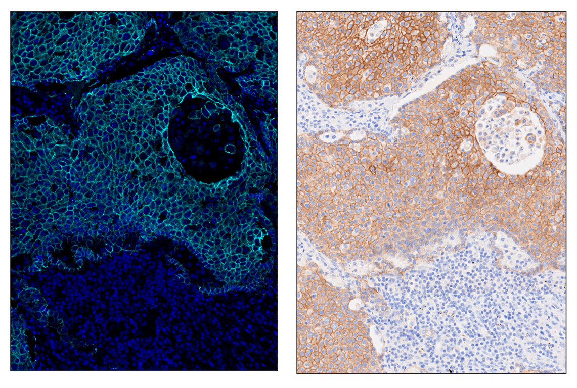
SignalStar™ multiplex immunohistochemical analysis of paraffin-embedded human squamous cell lung carcinoma using E-Cadherin (24E10) & CO-0103-750 SignalStar™ Oligo-Antibody Pair #65506 (left, cyan) and DAPI #4083 (left, blue) compared to chromogenic immunohistochemical analysis of a serial section of paraffin-embedded human squamous cell lung carcinoma using E-Cadherin (24E10) Rabbit mAb #3195 (right). All fluorophores have been assigned a pseudocolor, as indicated. Staining was performed on the BOND RX autostainer by Leica Biosystems.
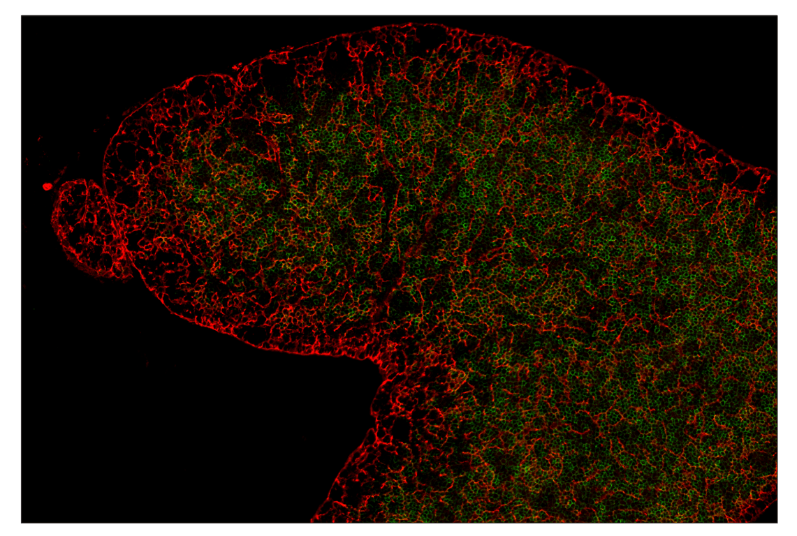
SignalStar™ multiplex immunohistochemical analysis of paraffin-embedded mouse thymus using CD28 (D2Z4E) & CO-0081-488 SignalStar™ Oligo-Antibody Pair #52935 (green), and E-Cadherin (24E10) & CO-0103-647 SignalStar™ Oligo-Antibody Pair #31774 (red). All fluorophores have been assigned a pseudocolor, as indicated. Staining was performed on the BOND RX autostainer by Leica Biosystems.