


 全部商品分类
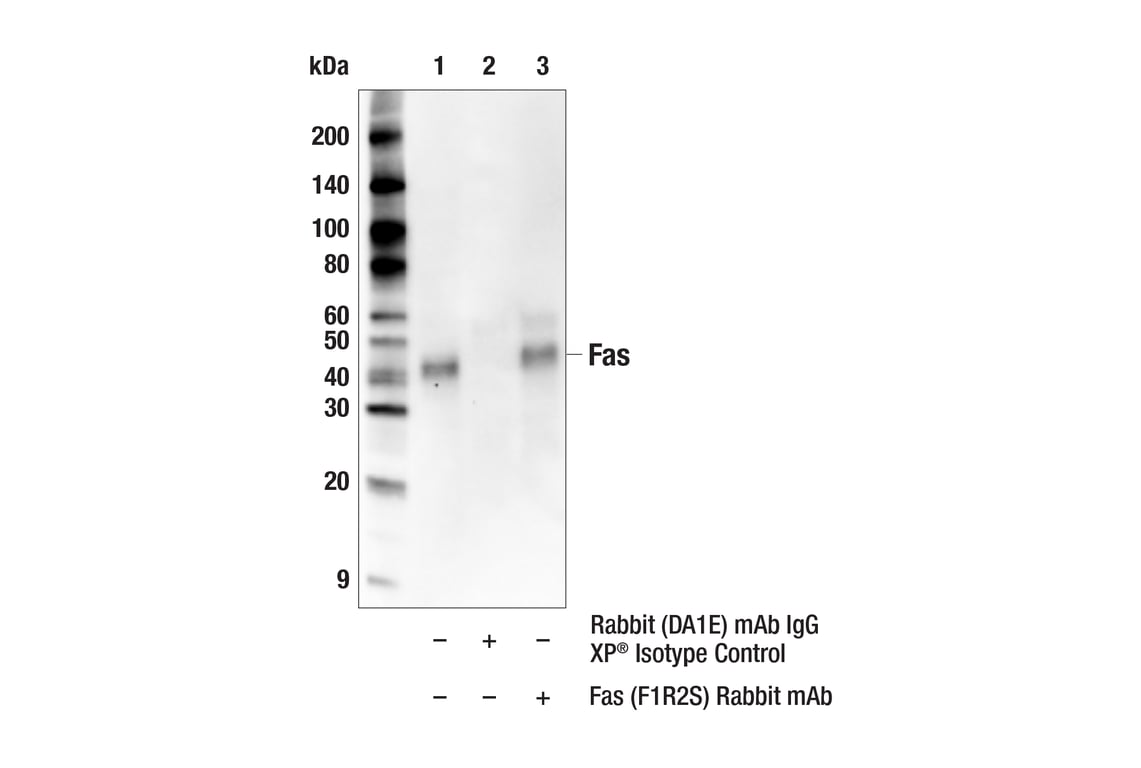


 用小程序,查商品更便捷


Monoclonal antibody is produced by immunizing animals with a synthetic peptide corresponding to residues surrounding Glu46 of mouse Fas protein, within the extracellular region.

Product Usage Information
| Application | Dilution |
|---|---|
| Western Blotting | 1:1000 |
| Simple Western™ | 1:10 - 1:50 |
| Immunoprecipitation | 1:50 |
Specificity/Sensitivity
Species Reactivity:
Mouse
Supplied in 10 mM sodium HEPES (pH 7.5), 150 mM NaCl, 100 µg/mL BSA, 50% glycerol, and less than 0.02% sodium azide. Store at –20°C. Do not aliquot the antibody.
参考图片
Immunoprecipitation of Fas protein from A20 cell extracts. Lane 1 is 10% input, lane 2 is Rabbit (DA1E) mAb IgG XP® Isotype Control #3900, and lane 3 is Fas (F1R2S) Rabbit mAb. Western blot analysis was performed using Fas (F1R2S) Rabbit mAb. Mouse Anti-rabbit IgG (Conformation Specific) (L27A9) mAb (HRP Conjugate) #5127 was used as a secondary antibody.
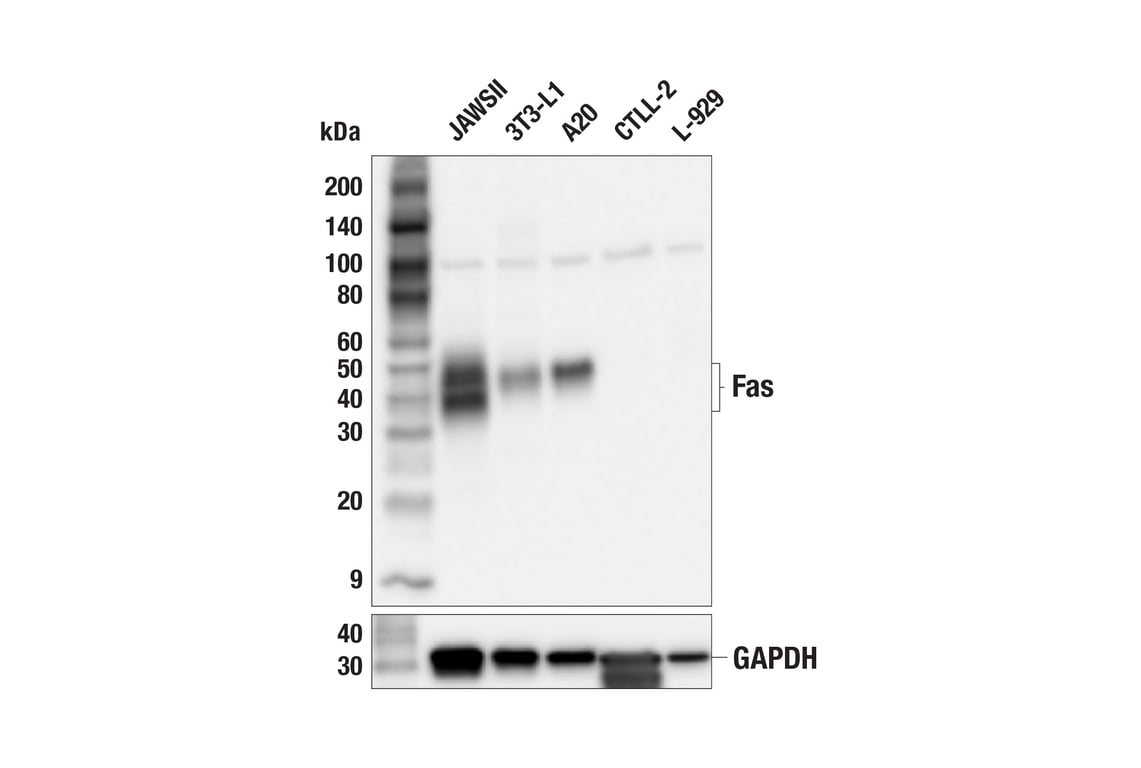
Western blot analysis of extracts from various cell lines using Fas (F1R2S) Rabbit mAb (upper) or GAPDH (D16H11) XP® Rabbit mAb #5174 (lower). Low expression of Fas protein in CTLL-2 and L-929 cells is consistent with published observations.
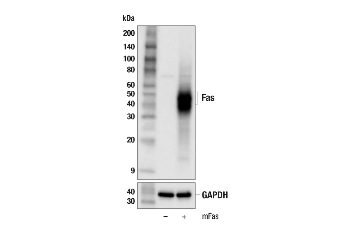
Western blot analysis of extracts from 293T cells, mock transfected (-) or transfected with a construct expressing full-length mouse Fas protein (mFas; +), using Fas (F1R2S) Rabbit mAb (upper) or GAPDH (D16H11) XP® Rabbit mAb #5174 (lower).
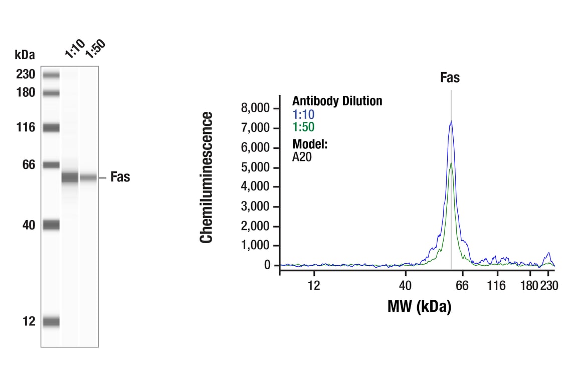
Simple WesternTM analysis of lysates (0.1 mg/mL) from A20 cells using Fas (F1R2S) Rabbit mAb #87871. The virtual lane view (left) shows the target band (as indicated) at 1:10 and 1:50 dilutions of primary antibody. The corresponding electropherogram view (right) plots chemiluminescence by molecular weight along the capillary at 1:10 (blue line) and 1:50 (green line) dilutions of primary antibody. This experiment was performed under reducing conditions on the JessTM Simple Western instrument from ProteinSimple, a BioTechne brand, using the 12-230 kDa separation module.