


 全部商品分类
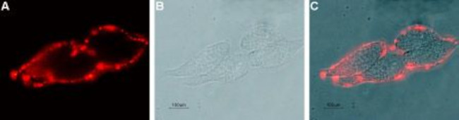


KCNN4 (KCA3.1- SK4) (EXTRACELL

 下载产品说明书
 用小程序,查商品更便捷


 收藏
收藏
 对比
对比 咨询
咨询种属反应
宿主/亚型
分类
类型
克隆号
抗原
偶联物
形式
浓度
纯化类型
保存液
内含物
保存条件
运输条件
产品详细信息
Reconstitution: 25 µL or 100 µL double distilled water (DDW), depending on the sample size. The antibody ships as a lyophilized powder at room temperature. Upon arrival, it should be stored at -20C. The reconstituted solution can be stored at 4C for up to 1 week. For longer periods, small aliquots should be stored at -20C. Avoid multiple freezing and thawing. Centrifuge all antibody preparations before use (10000 x g 5 min).
靶标信息
The protein encoded by this gene is part of a potentially heterotetrameric voltage-independent potassium channel that is activated by intracellular calcium. Activation is followed by membrane hyperpolarization, which promotes calcium influx. The encoded protein may be part of the predominant calcium-activated potassium channel in T-lymphocytes. This gene is similar to other KCNN family potassium channel genes, but it differs enough to possibly be considered as part of a new subfamily.
仅用于科研。不用于诊断过程。未经明确授权不得转售。
生物信息学
蛋白别名: Gardos channel; HIK1; hKCa4; IK1; IKCa; IKCa1; Intermediate conductance calcium-activated potassium channel protein 4; intermediate conductance K channel; intermediate-conductance Ca-activated K channel; KCa3.1; KCa4; KCNN 4; mIK1; mKCa4; potassium channel, calcium activated intermediate/small conductance subfamily N alpha, member 4; potassium intermediate/small conductance calcium-activated channel, subfamily N, member 4; putative erythrocyte intermediate conductance calcium-activated potassium Gardos channel; Putative Gardos channel; SK4; SKCa 4; SKCa4; small conductance calcium-activated potassium channel 4
基因别名: DHS2; hIKCa1; hKCa4; hSK4; IK; IK1; IKCA1; KCa3.1; KCA4; KCNN4; mIKCa1; rKCNN4c; rSK4; SK4; SKCas
UniProt ID:(Human) O15554, (Mouse) O89109
Entrez Gene ID:(Human) 3783, (Rat) 65206, (Mouse) 16534
参考图片
Expression of KCNN4in live intact human LN-CaP prostate carcinoma cells - Cell surface detection of KCNN4 in live intact human LN-CaP prostate carcinoma cells with Mouse Anti-KCNN4 (KCa3.1, SK4) (extracellular) Antibody (#ALM-051) (1:20), followed by goat- Anti-mouse-DyLight-594 secondary Antibody (red) (A). B. Live view of the cells. C. Merge of the two images.
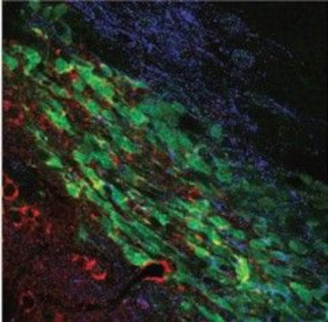
Multiplex staining of KCa3.1 (SK4) and TRPC1 in mouse brain - Immunohistochemical staining of mouse brain sections using Mouse Anti-KCNN4 (KCa3.1, SK4) (extracellular) Antibody (#ALM-051) and Anti-TRPC1 Antibody (#ACC-010). TRPC1 staining (blue) is detected in neuroblasts and outside neuroblasts as well (in astrocytes). KCa3.1 (red) is detected in neuroblasts. Merged image demonstrates the partial co-localization between the two. Adapted from Turner, K.L.et al. (2014)Cereb. Cortex.24,2388. with permissionof Oxford University Press.
Western blot analysis using Mouse Anti-KCNN4 (KCa3.1, SK4) (extracellular) Antibody (#ALM-051), (1:250): - 1. Mouse MS1 endothelial cells. 2. Rat IEC-6 intestinal epithelial cells. 3. Human LN-CaP prostate carcinoma cells. 4. Human THP-1 acute monocytic leukemia cells.
Western blot analysis using Mouse Anti-KCNN4 (KCa3.1, SK4) (extracellular) Antibody (#ALM-051), (1:250): - 1. HEK cells transfected with control vector. 2. HEK cells transfected with human KCa3.1.
Cell surface detection of KCa3.1 (SK4) in live intact THP-1 (human acute monocytic leukemia cells) cell line: - (black) Cells + Goat- Anti-mouse-Cy5. (red) Cells + Mouse IgM isotype control+ Goat- Anti-mouse-Cy5. (green) Cells + Mouse Anti-KCNN4 (KCa3.1, SK4) (extracellular) Antibody (#ALM-051), (1:20) + Goat- Anti-mouse-Cy5.
Cell surface detection of KCa3.1 (SK4) in live intact Raji (human Burkitts lymphoma B cells) cell line: - (black) Cells + goat- Anti-mouse-Cy5. (red) Cells + mouse IgM isotype control + goat- Anti-mouse-Cy5. (green) Cells + Mouse Anti-KCNN4 (KCa3.1, SK4) (extracellular) Antibody (#ALM-051), (1:20) + goat- Anti-mouse-Cy5.