


 全部商品分类
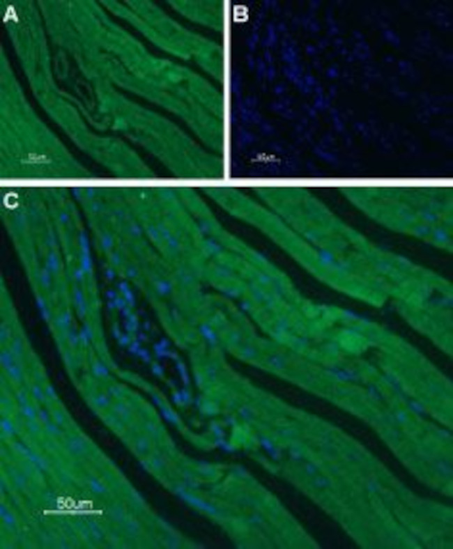


 下载产品说明书 下载SDS
 用小程序,查商品更便捷


 收藏
收藏
 对比
对比 咨询
咨询种属反应
宿主/亚型
分类
类型
抗原
偶联物
形式
浓度
纯化类型
保存液
内含物
保存条件
运输条件
产品详细信息
Reconstitution: 25 µL, 50 µL or 0.2 mL double distilled water (DDW), depending on the sample size. The antibody ships as a lyophilized powder at room temperature. Upon arrival, it should be stored at -20C. The reconstituted solution can be stored at 4C, protected from the light, for up to 1 week. For longer periods, small aliquots should be stored at -20C. Avoid multiple freezing and thawing. Centrifuge all antibody preparations before use (10000 X g 5 min).
靶标信息
CACNA1C encodes an alpha-1 subunit of a voltage-dependent calcium channel. Calcium channels mediate the influx of calcium ions into the cell upon membrane polarization. The alpha-1 subunit consists of 24 transmembrane segments and forms the pore through which ions pass into the cell. The calcium channel consists of a complex of alpha-1, alpha-2/delta, beta, and gamma subunits in a 1:1:1:1 ratio. There are multiple isoforms of each of these proteins, either encoded by different genes or the result of alternative splicing of transcripts. The protein encoded by CACNA1C binds to and is inhibited by dihydropyridine. Alternative splicing results in many transcript variants encoding different proteins.
仅用于科研。不用于诊断过程。未经明确授权不得转售。
生物信息学
蛋白别名: brain class C; CACH3; CACN4; CACNA 1D; CACNL1A2; calcium channel voltage-dependent alpha1c subunit; calcium channel, cardic dihydropyridine-sensitive, alpha-1 subunit; Calcium channel, L type, alpha-1 polypeptide, isoform 1, cardiac muscle; calcium channel, voltage-dependent, alpha 1C subunit; calcium channel, voltage-dependent, L type, alpha 1C subunit; DHP receptor; DHPR, alpha-1 subunit; L-type calcium channel alpha-1 subunit; long-NT; LTCC; MBC; MELC-CC; MGC120730; Mouse brain class C; neuronal voltage-gated calcium channel alpha 1C subunit; Rat brain class C; RBC; skeletal muscle-specific calcium channel; voltage-dependent L-type Ca2+ channel alpha 1 subunit; Voltage-dependent L-type calcium channel subunit alpha-1C; Voltage-gated calcium channel subunit alpha Cav1.2; voltage-gated L-type calcium channel Cav1.2 alpha 1 subunit, splice variant 10*
基因别名: CACH2; CACN2; CACNA1C; CACNL1A1; CaV1.2; CCHL1A1; D930026N18Rik; LQT8; MBC; MELC-CC; RATIVS302; TS
UniProt ID:(Human) Q13936, (Mouse) Q01815, (Rat) P22002
Entrez Gene ID:(Human) 775, (Mouse) 12288, (Rat) 24239
参考图片
Expression of CaV1.2 in rat heart - Immunohistochemical staining of rat heart paraffin embedded sections using Guinea pig Anti-CaV1.2 (CACNA1C) Antibody (#ACC-003-GP). A. CaV1.2 staining (green) appears mainly in the cardiac muscle, and in a lesser intensity in the tunica intima layer of the smooth muscle of the muscular arteries. B. Nuclear staining using DAPI as the counter stain. C. Merged images of A and B.
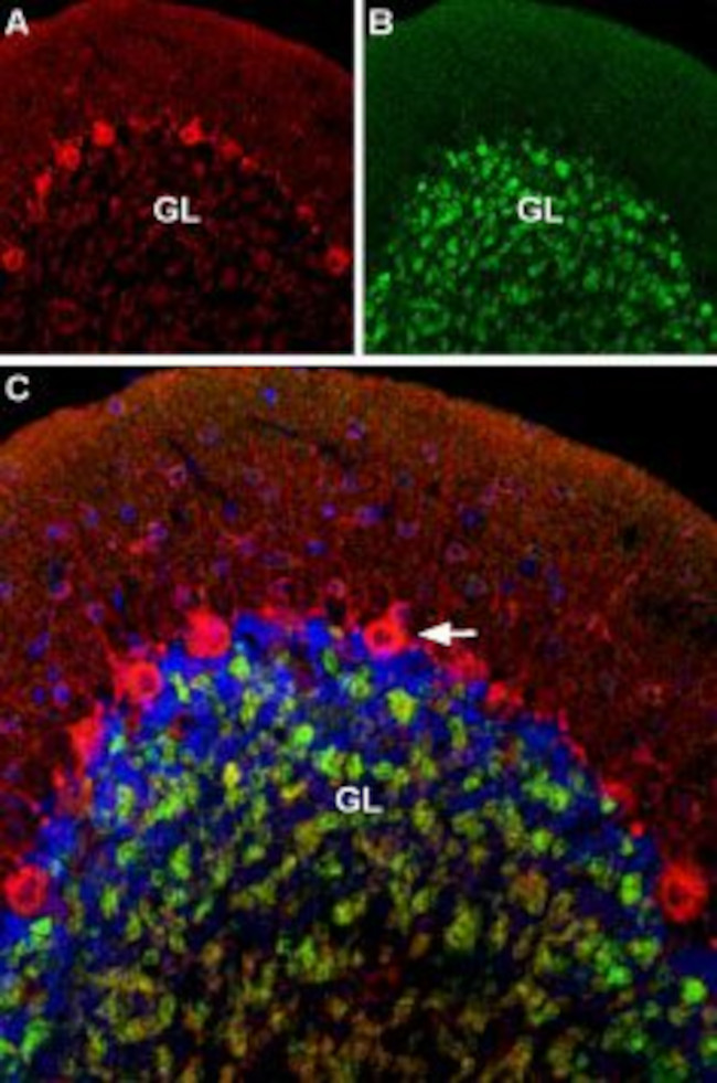
Multiplex staining of CaV1.2 and GABA (A) alpha 1 Receptor in rat cerebellum - Immunohistochemical staining of rat cerebellum using Guinea pig Anti-CaV1.2 (CACNA1C) Antibody (#ACC-003-GP) and Anti-GABA (A) alpha 1 Receptor (extracellular)-ATTO Fluor-488 Antibody (#AGA-001-AG).A. CaV1.2 (red) is detected mostly in Purkinje cells (arrow). B. In the same section,GABA (A) alpha 1 Receptor (green) is observed in the granule layer. C. Merge of the two images suggests some colocalization between CaV1.2 and GABA (A) alpha 1 Receptor in the rat granule layer but only CaV1.2 appears in Purkinje cells.
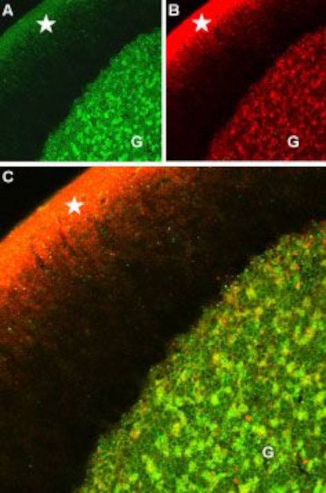
Multiplex staining of CaV1.2 and GABA (A) alpha 1 Receptor in rat cerebellum - Immunohistochemical staining of rat cerebellum using Guinea pig Anti-CaV1.2 (CACNA1C) Antibody (#ACC-003-GP) and Anti-GABA (A) alpha 1 Receptor (extracellular) Antibody (#AGA-001). A. CaV1.2 (green) is detected in the granule layer of the cerebellum (G) and in the upper molecular layer (star). B. In the same section,GABA (A) alpha 1 Receptor (red) is seen in the granule layer. C. Merge of the two images reveals high degree of colocalization between CaV1.2 and GABA (A) alpha 1 Receptor in the granule layer.
Expression of CaV1.2 in human atria - Immunohistochemical staining of human left atrium using Guinea pig Anti-CaV1.2 (CACNA1C) Antibody (#ACC-003-GP), (1:100).The picture was kindly provided by Dr. Van Wagoner, D.R. from the Department of Molecular Cardiology, Cleveland Clinic, Cleveland, Ohio, USA. Lovano, B. and Peterson, J. collected the data.
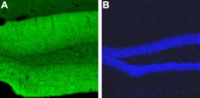
Expression of CaV1.2 in mouse hippocampus - Immunohistochemical staining of mouse dentate gyrus using Guinea pig Anti-CaV1.2 (CACNA1C) Antibody (#ACC-003-GP). A. CaV1.2 (green) appeared in the outer molecular layer of the dentate gyrus and in the granule layer. B. Counterstain with DAPI (blue) outlines the granule layer of the dentate gyrus.
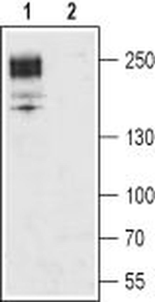
Western blot analysis of rat brain membrane: - 1.Guinea pig Anti-CaV1.2 (CACNA1C) Antibody (#ACC-003-GP), (1:200). 2. Guinea pig Anti-CaV1.2 (CACNA1C) Antibody , preincubated with Cav1.2/CACNA1C Blocking Peptide (#BLP-CC003).
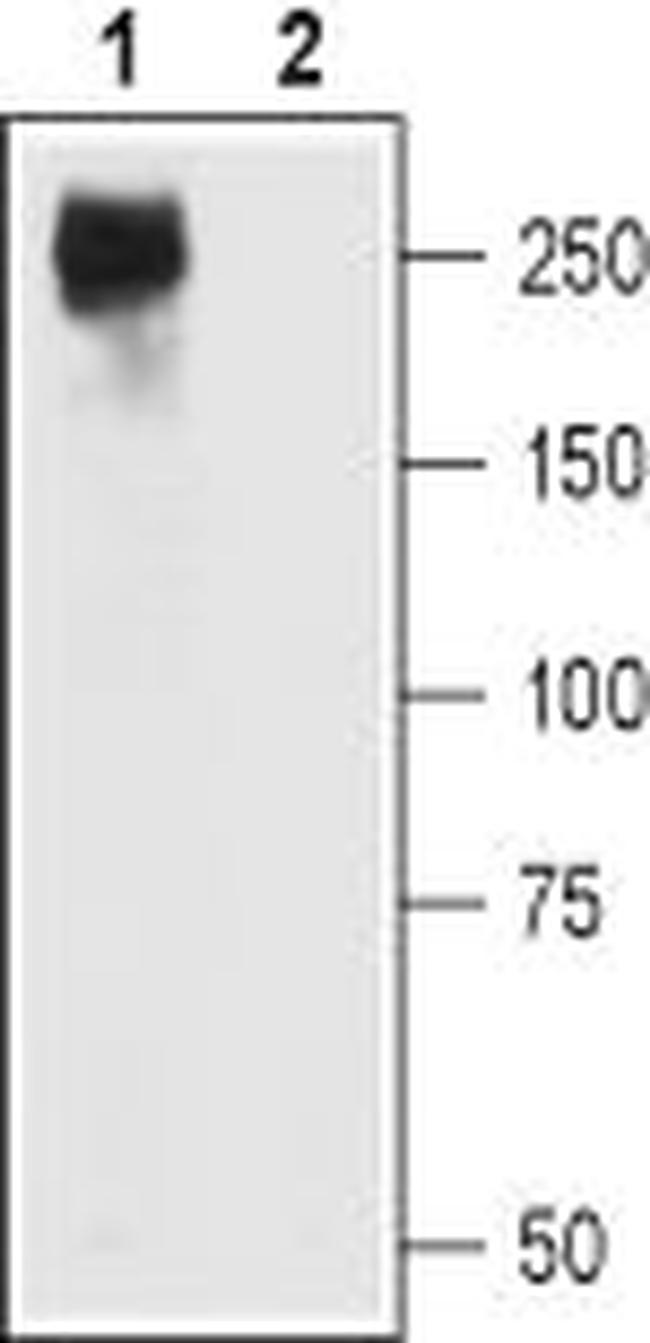
Western blot analysis of CaV1.2-transfectedXenopusoocytes (lane 1) and non-transfected oocytes lysates (lane 2): - 1.Guinea pig Anti-CaV1.2 (CACNA1C) Antibody (#ACC-003-GP), (1:200) inCaV1.2 (CACNA1C) Channel Overexpressed inXenopusoocytes. 2. Guinea pig Anti-CaV1.2 (CACNA1C) Antibody in non-transfected oocytes.