


 全部商品分类
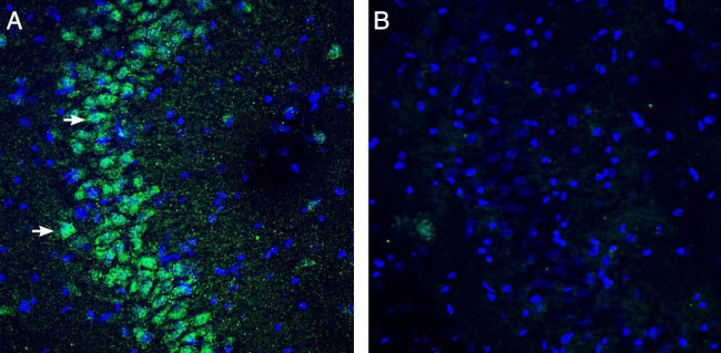


SLO BETA 3 (KCNMB3) (EXTRACELL

 下载产品说明书 下载SDS
 用小程序,查商品更便捷


 收藏
收藏
 对比
对比 咨询
咨询种属反应
宿主/亚型
分类
类型
抗原
偶联物
形式
浓度
纯化类型
保存液
内含物
保存条件
运输条件
产品详细信息
Reconstitution: 50 µL or 0.2 mL double distilled water (DDW), depending on the sample size. The antibody ships as a lyophilized powder at room temperature. Upon arrival, it should be stored at -20C. The reconstituted solution can be stored at 4C for up to 1 week. For longer periods, small aliquots should be stored at -20C. Avoid multiple freezing and thawing. Centrifuge all antibody preparations before use (10000 x g 5 min).
靶标信息
MaxiK channels are large conductance, voltage and calcium-sensitive potassium channels which are fundamental to the control of smooth muscle tone and neuronal excitability. MaxiK channels can be formed by 2 subunits: the pore-forming alpha subunit and the modulatory beta subunit. The protein encoded by this gene is an auxiliary beta subunit which may partially inactivate or slightly decrease the activation time of MaxiK alpha subunit currents. Alternative splicing results in multiple transcript variants. A related pseudogene has been identified on chromosome 22.
仅用于科研。不用于诊断过程。未经明确授权不得转售。
生物信息学
蛋白别名: BK beta 3; Calcium-activated potassium channel subfamily M subunit beta-3; Calcium-activated potassium channel subunit beta-3; kcnmb3 {ECO:0000312|EMBL:BAF79924.1}; Maxi K channel subunit beta-3; potassium large conductance calcium-activated channel, subfamily M beta member 3; potassium large conductance calcium-activated channel, subfamily M, beta member 4; slobeta3 (KCNMB3)
基因别名: EG435726; Gm5707; Kcnmb3
UniProt ID:(Rat) A7VL23
Entrez Gene ID:(Mouse) 100502876, (Rat) 310303
参考图片
Expression of KCNMB3 in rat hippocampus. Immunohistochemical staining of perfusion-fixed frozen rat brain sections with Anti-slo beta 3 (KCNMB3) (extracellular) Antibody (#APC-068), (1:300), followed by goat Anti-rabbit-Alexa-488. A. Staining in the hippocampal CA3 region, showed immunoreactivity (green) in cortical neurons (arrows). B. Pre-incubation of the Antibody with slo beta 3 (KCNMB3) (extracellular) Blocking Peptide (BLP-PC068), suppressed staining. Cell nuclei are stained with DAPI (blue).
Western blot analysis of mouse brain lysates (lanes 1 and 3) and rat pancreas membranes (lanes 2 and 4): - 1,2. Anti-slo beta 3 (KCNMB3) (extracellular) Antibody (#APC-068), (1:1000).3,4. Anti-slo beta 3 (KCNMB3) (extracellular) Antibody , preincubated with slo beta 3/KCNMB3 (extracellular) Blocking Peptide (#BLP-PC068).