


 全部商品分类
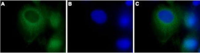


 下载产品说明书 下载SDS
 用小程序,查商品更便捷


 收藏
收藏
 对比
对比 咨询
咨询种属反应
宿主/亚型
分类
类型
抗原
偶联物
形式
浓度
纯化类型
保存液
内含物
保存条件
运输条件
产品详细信息
Reconstitution: 25 µL, 50 µL or 0.2 mL double distilled water (DDW), depending on the sample size. The antibody ships as a lyophilized powder at room temperature. Upon arrival, it should be stored at -20C. The reconstituted solution can be stored at 4C for up to 1 week. For longer periods, small aliquots should be stored at -20C. Avoid multiple freezing and thawing. Centrifuge all antibody preparations before use (10000 x g 5 min).
靶标信息
The mammalian transient receptor potential (TRP) superfamily can be divided into three major families including the "canonical TRP" (TRPC) family. The seven members of this family share the activation through PLC-coupled receptors and have been suggested to be components of receptor-regulated cation channels in different cell types. Furthermore, the members of the TRPC3/6/7 subfamily can be activated by diacylglycerol analogs, suggesting a possible mechanism of activation of these channels by PLC-coupled receptors. TRPC3 encodes a Ca2+-permeant channel that is agonist-activated but not store-operated or directly receptor-activated. TRPC3 physically interacts with TRPC6 and TRPC7 and forms functional tetrameric channels.
仅用于科研。不用于诊断过程。未经明确授权不得转售。
生物信息学
蛋白别名: hTrp-3; hTrp3; ion channel protein; mTrp3; Receptor-activated cation channel TRP3; Short transient receptor potential channel 3; transient receptor potential cation channel subfamily C member 3; transient receptor potential cation channel subfamily C member 3 variant c; transient receptor potential cation channel, subfamily C, member 3; Transient receptor protein 3; TRP-3; Trp-related protein 3; TrpC3
基因别名: Mwk; SCA41; Trcp3; TRP3; TRPC3; TrpC3c; Trrp3
UniProt ID:(Human) Q13507, (Mouse) Q9QZC1
Entrez Gene ID:(Human) 7222, (Rat) 60395, (Mouse) 22065
参考图片
Expression of TRPC3 in rat C6 brain glioma cells - Immunocytochemical staining of Paraformaldehyde-fixed and permeabilized rat C6 brain glioma cells. A. Staining using Anti-TRPC3 Antibody (#ACC-016), (1:500) followed by goat Anti-rabbit-AlexaFluor-488 secondary Antibody . B. Nuclear staining using the cell-permeable dye Hoechst 33342. C. Merged image of panels A and B.
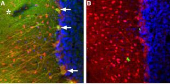
Expression of TRPC3 in rat cerebellum - Immunohistochemical staining of TRPC3 in rat cerebellum using Anti-TRPC3 Antibody (#ACC-016). A. TRPC3 (green) appears in Purkinje cells (arrows) including both soma and dendrites and as well as in the molecular layer neuropil (asterisk). Staining of the same section with mouse Anti-parvalbumin (red) reveals that TRPC3 is not expressed in molecular layer interneurons. B. Pre-incubating Anti-TRPC3 with the TRPC3 peptide antigen blocks staining. DAPI is used as the counterstain (blue).
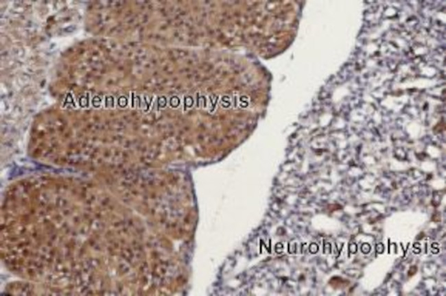
Expression of TRPC3 in rat pituitary gland - Immunohistochemical staining of rat pituitary gland paraffin embedded sections using Anti-TRPC3 Antibody (#ACC-016), (1:100). TRPC3 is mainly expressed in the adenohypophysis (on left). Hematoxilin is used as the counterstain.
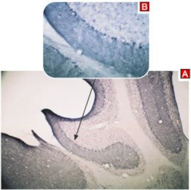
Expression of TRPC3 in mouse cerebellum - Immunohistochemical staining of mouse cerebellum using Anti-TRPC3 Antibody (#ACC-016) (A). Immunoreactivity appears in the molecular layer and in Purkinje cells (B).
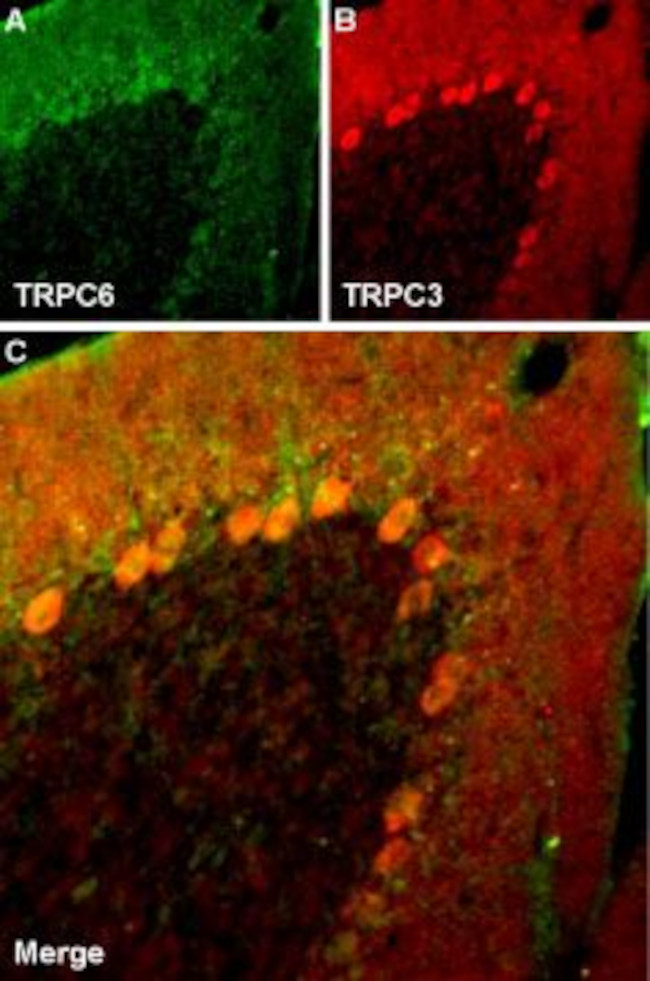
Multiplex staining of TRPC6 and TRPC3 in rat cerebellum - Immunohistochemical staining of rat cerebellum frozen section using Guinea pig Anti-TRPC6 Antibody (#ACC-017-GP) and rabbit Anti-TRPC3 Antibody (#ACC-016). A. TRPC6 staining (green) appears in molecular layer and in Purkinje cells. B. In the same section, staining of TRPC3 (red) appears as well in both molecular layer and Purkinje cells. C. Merge images of A and B indicates extensive co-localization. DAPI is used as the counterstain (blue).
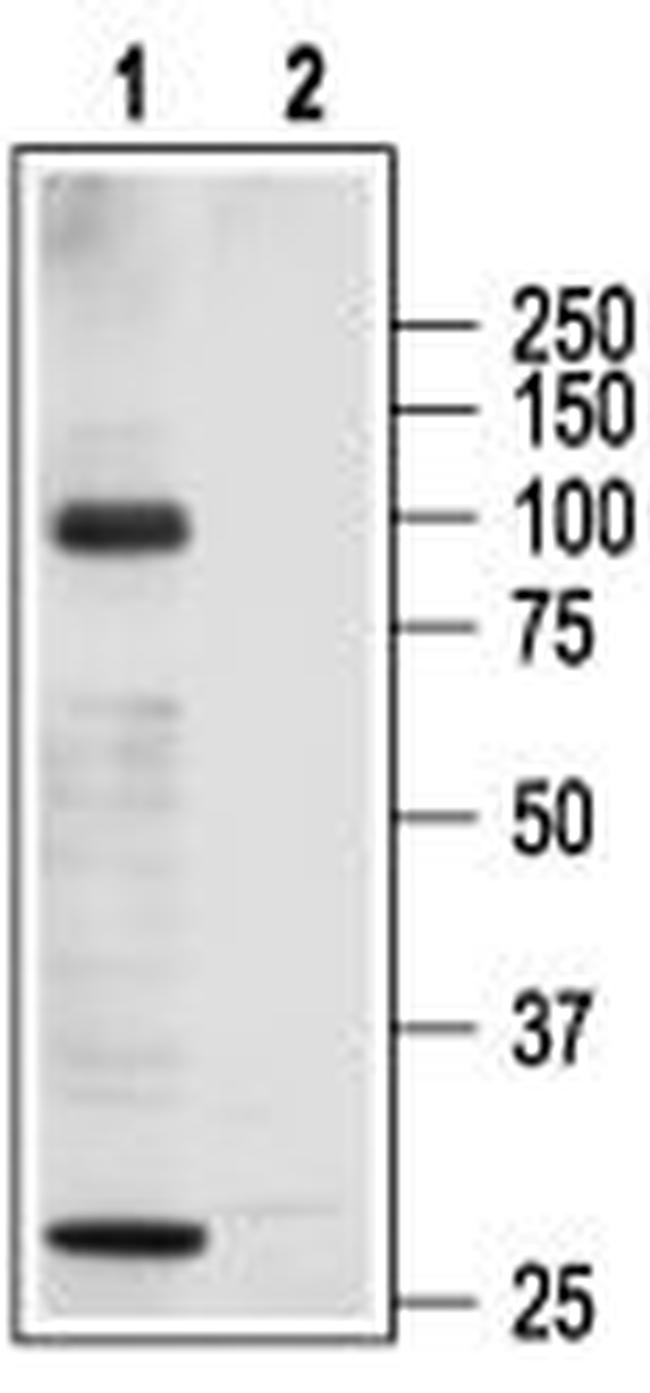
Western blotanalysisof rat heart membranes: - 1. Anti-TRPC3 Antibody (#ACC-016), (1:200). 2. Anti-TRPC3 Antibody , preincubated with TRPC3 Blocking Peptide (#BLP-CC016).