


 全部商品分类


NMDAR1 (GLUN1) (EXTRACELLULAR)

 下载产品说明书 下载SDS
 用小程序,查商品更便捷


 收藏
收藏
 对比
对比 咨询
咨询种属反应
宿主/亚型
分类
类型
抗原
偶联物
形式
浓度
纯化类型
保存液
内含物
保存条件
运输条件
产品详细信息
Reconstitution: 25 µL, 50 µL or 0.2 mL double distilled water (DDW), depending on the sample size. The antibody ships as a lyophilized powder at room temperature. Upon arrival, it should be stored at -20C. The reconstituted solution can be stored at 4C for up to 1 week. For longer periods, small aliquots should be stored at -20C. Avoid multiple freezing and thawing. Centrifuge all antibody preparations before use (10000 x g 5 min).
靶标信息
NMDAR1 encodes a protein that is a critical subunit of N-methyl-D-aspartate receptors, members of the glutamate receptor channel superfamily which are heteromeric protein complexes with multiple subunits arranged to form a ligand-gated ion channel. These subunits play a key role in the plasticity of synapses, which is believed to underlie memory and learning. Cell-specific factors are thought to control expression of different isoforms, possibly contributing to the functional diversity of the subunits. Alternatively spliced transcript variants have been described.
仅用于科研。不用于诊断过程。未经明确授权不得转售。
生物信息学
蛋白别名: GluN1; Glun1r; glutamate [NMDA] receptor subunit zeta 1; Glutamate [NMDA] receptor subunit zeta-1; glutamate receptor; Glutamate receptor ionotropic, NMDA 1; glutamate receptor, ionotropic, N-methyl D-aspartate 1; N-methyl-D-aspartate glutamate receptor; N-methyl-D-aspartate receptor channel, subunit zeta-1; N-methyl-D-aspartate receptor subunit NR1; neurotransmitter receptor; NMD-R1; NMDA R1 receptor C1 cassette; NMDA receptor 1; NMDZ1; NMZ1; NR1C1; NR1C2; NR1C2'; NR1N1; pGluN1; RP11-350O14.1
基因别名: GluN1; GluRdelta1; Glurz1; GluRzeta1; GRIN1; M100174; MRD8; NMD-R1; NMDA1; Nmdar; NMDAR1; NR1; Rgsc174
UniProt ID:(Human) Q05586, (Rat) P35439, (Mouse) P35438
Entrez Gene ID:(Human) 2902, (Rat) 24408, (Mouse) 14810
参考图片
Multiplex staining of NMDAR1 and CALHM1 in mouse hippocampal CA1 region - Immunohistochemical staining of perfusion-fixed frozen mouse brain sections using Anti-NMDAR1 (GluN1) (extracellular) Antibody (#AGC-001), (1:200) and Anti-CALHM1-ATTO Fluor-594 Antibody (#ACC-101-AR), (1:60). A. Sections were stained with Anti-NMDAR1 (GluN1) (extracellular) Antibody , followed by goat- Anti-rabbit-Cy2 (green). Staining reveals expression in neurons of the pyramidal layer (an arrow points at the layer). B. The same section was incubated with Anti-CALHM1-ATTO Fluor-594 Antibody (red). C. Merge of the two images demonstrates colocalization of NMDAR1 and CALHM1 in pyramidal neurons.
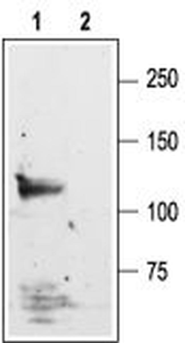
Western blot analysisof rat brain lysate: - 1. Anti-NMDAR1 (GluN1) (extracellular) Antibody (#AGC-001), (1:600). 2. Anti-NMDAR1 (GluN1) (extracellular) Antibody , preincubated with NMDAR1/GluN1 (extracellular) Blocking Peptide (#BLP-GC001).