


 全部商品分类
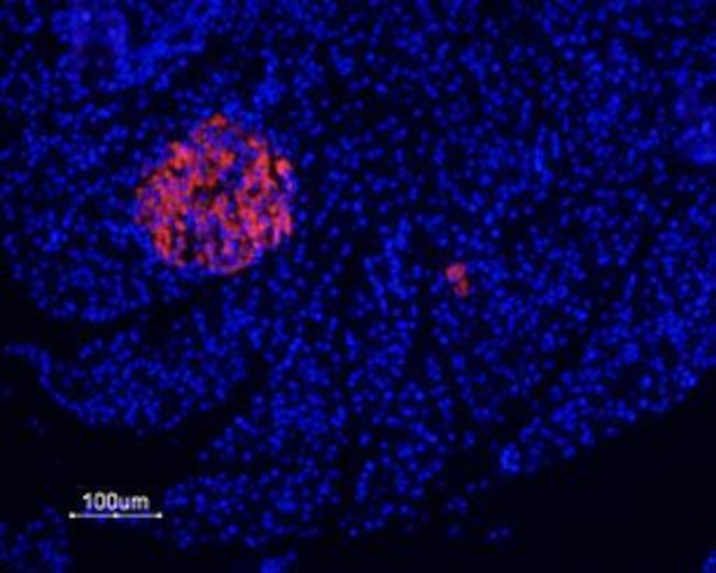


 下载产品说明书 下载SDS
 用小程序,查商品更便捷


 收藏
收藏
 对比
对比 咨询
咨询种属反应
宿主/亚型
分类
类型
抗原
偶联物
激发/发射光谱
形式
浓度
纯化类型
保存液
内含物
保存条件
运输条件
产品详细信息
Reconstitution: 50 µL double distilled water (DDW), depending on the sample size. The antibody ships as a lyophilized powder at room temperature. Upon arrival, it should be stored at -20C. The reconstituted solution can be stored at 4C for up to 1 week. For longer periods, small aliquots should be stored at -20C. Avoid multiple freezing and thawing. Centrifuge all antibody preparations before use (10000 x g 5 min).
靶标信息
The product of this gene belongs to the family of purinoceptors for ATP. This receptor functions as a ligand-gated ion channel and is responsible for ATP-dependent lysis of macrophages through the formation of membrane pores permeable to large molecules. Activation of this nuclear receptor by ATP in the cytoplasm may be a mechanism by which cellular activity can be coupled to changes in gene expression. Multiple alternatively spliced variants have been identified, most of which fit nonsense-mediated decay criteria.
仅用于科研。不用于诊断过程。未经明确授权不得转售。
生物信息学
蛋白别名: ATP receptor; MGC20089; P2X purinoceptor 7; p2X receptor 7; P2X7; P2X7 purinoceptor; P2X7 receptor; P2Z; P2Z receptor; Purinergic receptor; purinergic receptor P2X, ligand gated ion channel, 7; purinergic receptor P2X, ligand-gated ion channel, 7; purinergic receptor P2X7; purinergic receptor P2X7 variant A
基因别名: AI467586; P2RX7; P2X(7); P2X7; P2X7R
UniProt ID:(Human) Q99572, (Rat) Q64663, (Mouse) Q9Z1M0
Entrez Gene ID:(Human) 5027, (Rat) 29665, (Mouse) 18439
参考图片
Expression of P2X7 Receptorin rat pancreas - Immunohistochemical staining of rat paraffin embedded endocrine and exocrine pancreas sections using Anti-P2X7 Receptor-ATTO Fluor-550 Antibody (#APR-004-AO), (1:20), (red). Staining is highly specific for endocrine cells of the Isle of Langerhans. Hoechst 33342 is used as the counterstain (blue).
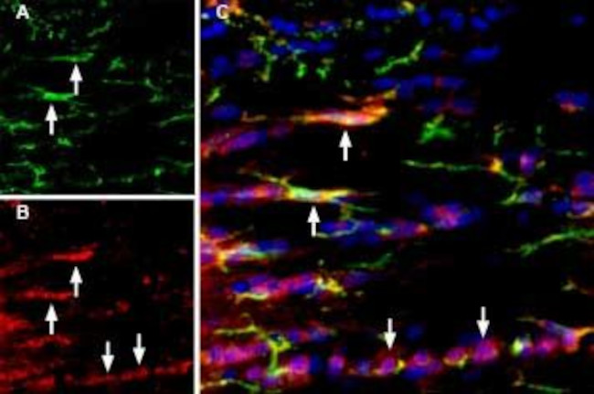
Multiplex staining of IBA1/AIF1 and P2X7 in rat brain. Immunohistochemical staining of rat corpus callosum (CC) free floating frozen sections using Anti-IBA1/AIF1 Antibody (#ACS-010), (1:1000) and Anti-P2X7 Receptor-ATTO Fluor-550 Antibody (#APR-004-AO) (1:60). A. IBA1/AIF1 immunoreactivity (green) appears in microglia (arrows). B. P2X7 immunostaining (red) appears in microglia (up-pointing arrows) and in other cell types in the corpus callosum (down-pointing arrows). C. Merged image of panels A and B demonstrates partial colocalization of both proteins. Nuclei are demonstrated using DAPI as the counterstain (blue).
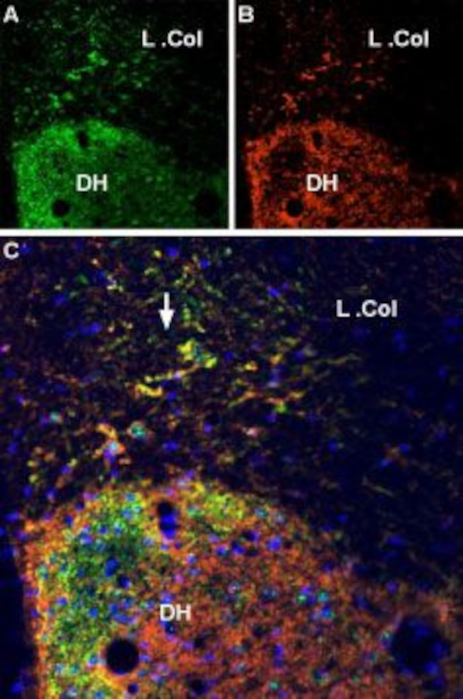
Multiplex staining of VGLUT2 and P2X7 Receptor in rat spinal cord - Immunohistochemical staining of perfusion-fixed frozen rat spinal cord sections using Anti-VGLUT2 Antibody (#AGC-036), (1:600) and Anti-P2X7 Receptor-ATTO Fluor-550 Antibody (#APR-004-AO), (1:100). A. Vesicular Glutamate Transporter 2 labeling followed by goat- Anti-rabbit-Alexa-488 (green). B. The same section labeled for P2X7 Receptor (orange). C. Merge of A and B demonstrates partial co-localization of VGLUT2 and P2X7 Receptor in dorsal horn and in lateral column (L. Col., arrow). Cell nuclei were stained with DAPI (blue).
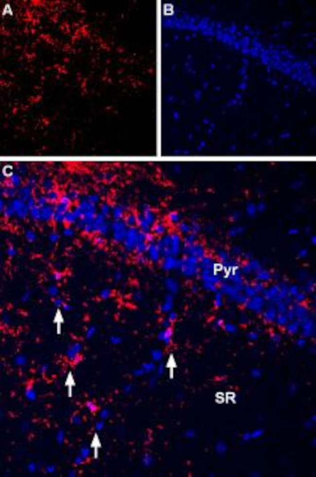
Expression ofP2RX7in rat hippocampus - Immunohistochemical staining of rat hippocampus frozen section using Anti-P2X7 Receptor-ATTO Fluor-550 Antibody (#APR-004-AO), (1:60). A. P2X7 staining (red) appears in the CA1 pyramidal (Pyr) and stratum radiatum (SR) layers in cells with glial morphology (arrows). B. DAPI is used as the counterstain (blue). C. Merged image of A and B.