


 全部商品分类
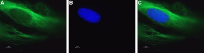


 下载产品说明书 下载SDS
 用小程序,查商品更便捷


 收藏
收藏
 对比
对比 咨询
咨询种属反应
宿主/亚型
分类
类型
抗原
偶联物
激发/发射光谱
形式
浓度
纯化类型
保存液
内含物
保存条件
运输条件
产品详细信息
Reconstitution: 50 µL double distilled water (DDW), depending on the sample size. The antibody ships as a lyophilized powder at room temperature. Upon arrival, it should be stored at -20C. The reconstituted solution can be stored at 4C for up to 1 week. For longer periods, small aliquots should be stored at -20C. Avoid multiple freezing and thawing. Centrifuge all antibody preparations before use (10000 x g 5 min).
靶标信息
CACNA1C encodes an alpha-1 subunit of a voltage-dependent calcium channel. Calcium channels mediate the influx of calcium ions into the cell upon membrane polarization. The alpha-1 subunit consists of 24 transmembrane segments and forms the pore through which ions pass into the cell. The calcium channel consists of a complex of alpha-1, alpha-2/delta, beta, and gamma subunits in a 1:1:1:1 ratio. There are multiple isoforms of each of these proteins, either encoded by different genes or the result of alternative splicing of transcripts. The protein encoded by CACNA1C binds to and is inhibited by dihydropyridine. Alternative splicing results in many transcript variants encoding different proteins.
仅用于科研。不用于诊断过程。未经明确授权不得转售。
生物信息学
蛋白别名: brain class C; CACH3; CACN4; CACNA 1D; CACNL1A2; calcium channel voltage-dependent alpha1c subunit; calcium channel, cardic dihydropyridine-sensitive, alpha-1 subunit; Calcium channel, L type, alpha-1 polypeptide, isoform 1, cardiac muscle; calcium channel, voltage-dependent, alpha 1C subunit; calcium channel, voltage-dependent, L type, alpha 1C subunit; DHP receptor; DHPR, alpha-1 subunit; L-type calcium channel alpha-1 subunit; long-NT; LTCC; MBC; MELC-CC; MGC120730; Mouse brain class C; neuronal voltage-gated calcium channel alpha 1C subunit; Rat brain class C; RBC; skeletal muscle-specific calcium channel; voltage-dependent L-type Ca2+ channel alpha 1 subunit; Voltage-dependent L-type calcium channel subunit alpha-1C; Voltage-gated calcium channel subunit alpha Cav1.2; voltage-gated L-type calcium channel Cav1.2 alpha 1 subunit, splice variant 10*
基因别名: CACH2; CACN2; CACNA1C; CACNL1A1; CaV1.2; CCHL1A1; D930026N18Rik; LQT8; MBC; MELC-CC; RATIVS302; TS
UniProt ID:(Human) Q13936, (Mouse) Q01815, (Rat) P22002
Entrez Gene ID:(Human) 775, (Mouse) 12288, (Rat) 24239
参考图片
Expression of CaV1.2 in mouse pancreatic microvascular endothelial cells (MS1) - Immunocytochemical staining of MS1 cells. A. Intracellular staining of paraformaldehyde-fixed and permeabilized MS1 cells with Anti-CaV1.2 (CACNA1C)-ATTO Fluor-488 Antibody (#ACC-003-AG), (1:50). B. The cell-permeable dye Hoechst 33342 (blue) was used for nuclear staining. C. Merged image of panels A and B.
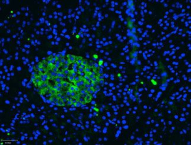
Expression of CaV1.2 in rat pancreas - Immunohistochemical staining of paraffin embedded sections of rat pancreas using Anti-CaV1.2 (CACNA1C)-ATTO Fluor-488 Antibody (#ACC-003-AG), (1:50). Staining is highly specific for endocrine cells of theIsle of Langerhans (IL).
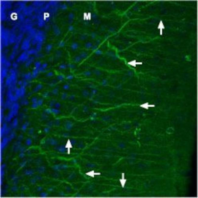
Expression of CaV1.2 in rat cerebellum - Immunohistochemical staining of rat cerebellum frozen sections with Anti-CaV1.2 (CACNA1C)-ATTO Fluor-488 Antibody (#ACC-003-AG), (1:100). Both dendrites of Purkinje cells (horizontal arrows) and fibers of Bergmann glia (vertical arrows) were stained. DAPI (blue Nissl counterstain) helps define the layers: granule (G), Purkinje (P) and molecular (M).