


 全部商品分类
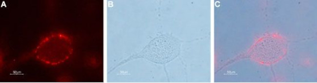


 下载产品说明书 下载SDS
 用小程序,查商品更便捷


 收藏
收藏
 对比
对比 咨询
咨询种属反应
宿主/亚型
分类
类型
抗原
偶联物
形式
浓度
纯化类型
保存液
内含物
保存条件
运输条件
产品详细信息
Reconstitution: 25 µL, 50 µL or 0.2 mL double distilled water (DDW), depending on the sample size. The antibody ships as a lyophilized powder at room temperature. Upon arrival, it should be stored at -20C. The reconstituted solution can be stored at 4C for up to 1 week. For longer periods, small aliquots should be stored at -20C. Avoid multiple freezing and thawing. Centrifuge all antibody preparations before use (10000 x g 5 min).
靶标信息
Gap junctions are channel-forming structures that allow direct metabolic and electrical communication between adjacent cells of almost all types in mammalian tissues. In the human body, they are absent only in adult skeletal muscle cells and some circulating blood cells. A gap junction is formed two hemichannels, one in each of the neighboring cells, composed of six subunits. In mice and humans, at least 20 connexin and 3 pannexin genes encode gap junction proteins. Connexins are only found in chordates, while pannexins are present in both chordate and invertebrate genomes. Pannexins, previously known as innexins, are predicted to have four transmembrane regions, two extracellular loops, one intracellular loop, and intracellular N- and C-termini. Both human and mouse genomes contain three pannexin-encoded genes. Pannexin 2 (Px2, PANX2) appears to be a brain specific gene, and is abundantly expressed in the central nervous system, as is pannexin 1. In many neuronal cell populations, including hippocampus, olfactory bulb, cortex, and cerebellum, pannexin 1 and pannexin 2 are co-expressed; in other brain regions such as white matter, only pannexin 1-positive cells are found.
仅用于科研。不用于诊断过程。未经明确授权不得转售。
生物信息学
蛋白别名: h PX2; Pannexin-2
基因别名: hPANX2; PANX2; PX2
UniProt ID:(Human) Q96RD6, (Mouse) Q6IMP4, (Rat) P60571
Entrez Gene ID:(Human) 56666, (Mouse) 406218, (Rat) 362979
参考图片
Expression of Pannexin 2 in human U-87 MG glioblastoma cell line - Cell surface detection of Pannexin 2 in live intact human U-87 MG glioblastoma cells. A. Cells were stained with Anti-Pannexin 2 (extracellular) Antibody (#ACC-232), (1:50), followed by goat Anti-rabbit-AlexaFluor-594 secondary Antibody (red). B. Live view of the cells. C. Merge of A and B.
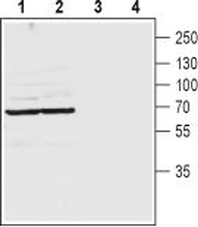
Western blot analysis of rat (lanes 1 and 3) and mouse (lanes 2 and 4) brain lysates: - 1,2. Anti-Pannexin 2 (extracellular) Antibody (#ACC-232), (1:200).3,4. Anti-Pannexin 2 (extracellular) Antibody , preincubated with Pannexin 2 (extracellular) Blocking Peptide (#BLP-CC232).