


 全部商品分类


NMDAR2A (GLUN2A) (EXTRACELLULA

 下载产品说明书
 用小程序,查商品更便捷


 收藏
收藏
 对比
对比 咨询
咨询种属反应
宿主/亚型
分类
类型
抗原
偶联物
形式
浓度
纯化类型
保存液
内含物
保存条件
运输条件
产品详细信息
Reconstitution: 25 µL, 50 µL or 0.2 mL double distilled water (DDW), depending on the sample size. The antibody ships as a lyophilized powder at room temperature. Upon arrival, it should be stored at -20C. The reconstituted solution can be stored at 4C for up to 1 week. For longer periods, small aliquots should be stored at -20C. Avoid multiple freezing and thawing. Centrifuge all antibody preparations before use (10000 x g 5 min).
靶标信息
N-methyl-D-aspartate (NMDA) receptors are a class of ionotropic glutamate-gated ion channels. These receptors have been shown to be involved in long-term potentiation, an activity-dependent increase in the efficiency of synaptic transmission thought to underlie certain kinds of memory and learning. NMDA receptor channels are heteromers composed of the key receptor subunit NMDAR1 (GRIN1) and 1 or more of the 4 NMDAR2 subunits: NMDAR2A (GRIN2A), NMDAR2B (GRIN2B), NMDAR2C (GRIN2C) and NMDAR2D (GRIN2D). Alternatively spliced transcript variants encoding different isoforms have been found for this gene.
仅用于科研。不用于诊断过程。未经明确授权不得转售。
生物信息学
蛋白别名: GluN2A; GluRepsilon1; Glutamate [NMDA] receptor subunit epsilon-1; glutamate receptor; Glutamate receptor ionotropic, NMDA 2A; glutamate receptor, ionotropic, N-methyl D-aspartate 2A; hNR2A; N-methyl D-aspartate receptor subtype 2A; N-methyl-D-aspartate receptor channel, subunit epsilon-1; N-methyl-D-aspartate receptor subunit 2A; NMDA Receptor 2A; NMDAR 2A; NMDAR2A; OTTHUMP00000160135; OTTHUMP00000174531
基因别名: EPND; FESD; GluN2A; GRIN2A; LKS; NMDAR2A; NR2A
UniProt ID:(Human) Q12879, (Mouse) P35436, (Rat) Q63728
Entrez Gene ID:(Human) 2903, (Mouse) 14811, (Rat) 24409
参考图片
Expression of NR2A in rat C6 glioma cells - Cell surface detection of NR2A in live intact rat C6 glioma cells. A. Cells werestained with Anti-NMDAR2A (GluN2A) (extracellular) Antibody (#AGC-002), (1:100), followed by goat Anti-rabbit-AlexaFluor-555 secondary Antibody (red). Cell nuclei were stained with the cell permeable dye Hoechst 33342 (blue staining). B. Live view of the same field.
Multiplex staining of GluN2A and GluN2B in mouse deep cerebellar nucleus - Immunohistochemical staining of perfusion-fixed frozen mouse brain sections using Anti-NMDAR2B (GluN2B) (extracellular)-ATTO Fluor-594 Antibody (#AGC-003-AR), (1:60) and Anti-NMDAR2A (GluN2A) (extracellular) Antibody (#AGC-002), (1:200). A. Sections were incubated with Anti-NMDAR2A (GluN2A) (extracellular) Antibody , followed by goat Anti-rabbit-Alexa-488 (green). B. The same sections were incubated with Anti-NMDAR2B (GluN2B) (extracellular)-ATTO Fluor-594 Antibody (red). C. Merge of A and B demonstrates the ubiquitous colocalization of the GluN2A and GluN2B subunits in cells with neuronal profiles in this nucleus. Arrows point at an example of NR2A and NR2B co-expression.
Expression of NR2A in rat hippocampus - Immunohistochemical staining of rat hippocampaldentate gyrus with Anti-NMDAR2A (GluN2A) (extracellular) Antibody (#AGC-002). A.NMDAR2A (green) appears diffusely in the outer molecular layer of thedentate gyrus (Out Mol.) and in cells along the subgranular layer (arrows). B. Staining of parvalbumin (PV, red) identifies interneurons in thedentate gyrus. C. Confocal merge demonstrates localization of PV in some neurons with NMDAR2A.
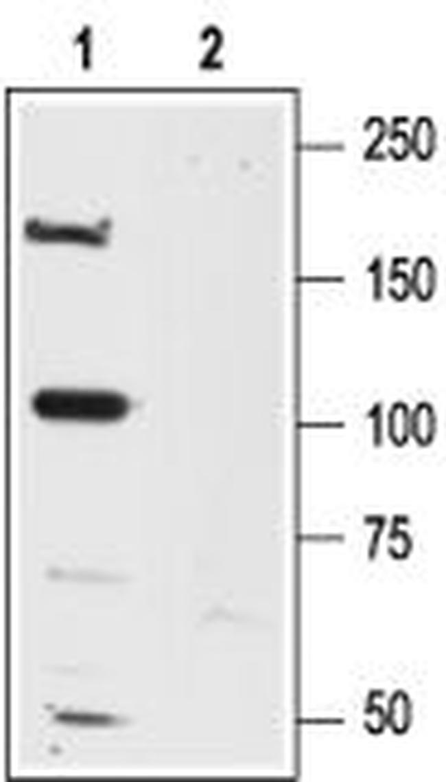
Western blot analysisof rat brain lysates: - 1. Anti-NMDAR2A (GluN2A) (extracellular) Antibody (#AGC-002), (1:600). 2. Anti-NMDAR2A (GluN2A) (extracellular) Antibody , preincubated with NMDAR2A/GluN2A (extracellular) Blocking Peptide (#BLP-GC002).
Immunoprecipitation of rat brain lysates - 1. Cell lysate. 2. Cell lysates + protein A beads + Anti-NMDAR2A (GluN2A) (extracellular) Antibody (#AGC-002).3. Cell lysates + protein A beads + pre-immune rabbit serum.Black arrow indicates the NR2A protein while the red arrow shows the IgG heavy chain. Immunoblot was performed with Anti-NMDAR2A (GluN2A) (extracellular) Antibody .