


 全部商品分类
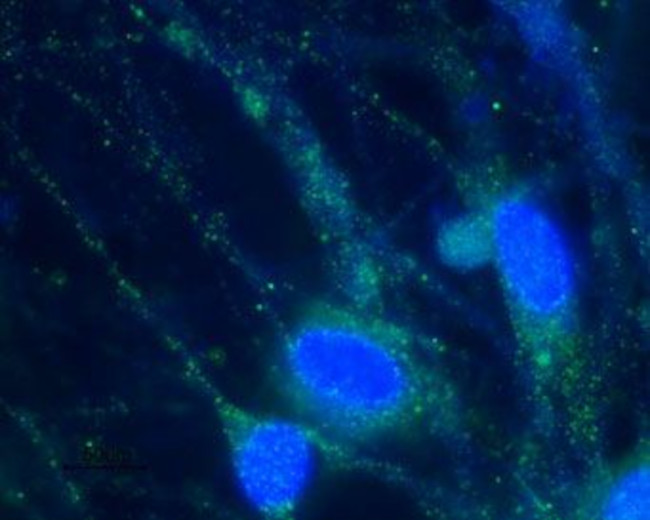


 下载产品说明书 下载SDS
 用小程序,查商品更便捷


 收藏
收藏
 对比
对比 咨询
咨询种属反应
宿主/亚型
分类
类型
抗原
偶联物
形式
浓度
纯化类型
保存液
内含物
保存条件
运输条件
产品详细信息
Reconstitution: 25 µL, 50 µL or 0.2 mL double distilled water (DDW), depending on the sample size. The antibody ships as a lyophilized powder at room temperature. Upon arrival, it should be stored at -20C. The reconstituted solution can be stored at 4C for up to 1 week. For longer periods, small aliquots should be stored at -20C. Avoid multiple freezing and thawing. Centrifuge all antibody preparations before use (10000 x g 5 min).
靶标信息
Hyperpolarization-activated cation channels of the HCN gene family such as HCN2, contribute to spontaneous rhythmic activity in both heart and brain. HCN2 is a member of a family of pacemaker channels activated by hyperpolarization and regulated by cyclic nucleotides. HCN1 and HCN2 play an important role for motor learning and neuronal integration by cerebellar Purkinje cells; as well as, shaping autonomous activity of single neurons and the periodicity of network oscillations. The HCN1 expression is highly enriched in cerebral cortex, hippocampus, cerebellum, and facial motor nucleus. HCN2 is highly abundant in mamillary bodies, pontine nucleus, ventral cochlear nucleus, and nucleus of the trapezoid body. These variations in regional specificity of HCN channels could generate important differences in neuronal pacemaker activity across brain systems.
仅用于科研。不用于诊断过程。未经明确授权不得转售。
生物信息学
蛋白别名: BCNG-2; brain cyclic nucleotide gated channel 2; Brain cyclic nucleotide-gated channel 2; HAC-1; hyperpolarization activated cyclic nucleotide-gated potassium channel 2; Hyperpolarization-activated cation channel 1; Potassium/sodium hyperpolarization-activated cyclic nucleotide-gated channel 2
基因别名: BCNG-2; BCNG2; HAC-1; HAC1; HCN2; trls
UniProt ID:(Human) Q9UL51, (Mouse) O88703, (Rat) Q9JKA9
Entrez Gene ID:(Human) 610, (Mouse) 15166, (Rat) 114244
参考图片
Expression of HCN2 in rat DRG primary culture - Immunocytochemical staining of paraformaldehyde-fixed and permeabilized rat dorsal root ganglion (DRG) primary culture using Anti-HCN2 Antibody (#APC-030), (1:100), (green). Cells were stained with Anti-HCN2 Antibody followed by goat Anti-rabbit-AlexaFluor-488 secondary Antibody . Nuclear staining of cells using the cell-permeable DNA dye Hoechst 33342 (blue).
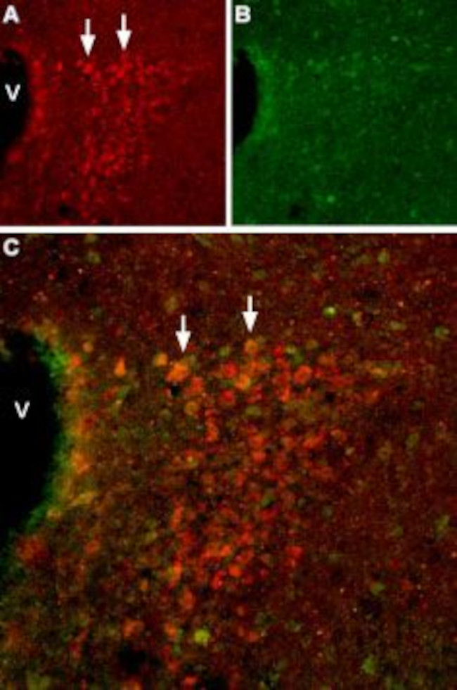
Expression of HCN2 in mousehypothalamus - Immunohistochemical staining of mouse hypothalamus using Anti-HCN2 Antibody (#APC-030). A. HCN2 (red) appears in cells of the paraventricular nucleus (PVN, arrows). B. Staining of paraventricular nerve cells with mouse Anti-calcium binding protein (CBD28k, green). C. Confocal merge of HCN2 and CBD28k demonstrates some co-localization. V = Third ventricle.
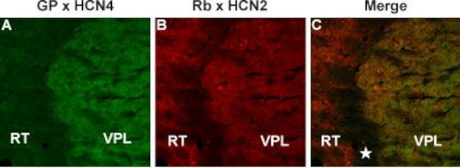
Colocalization of HCN4 and HCN2 in mouse thalamus - Immunohistochemical staining of mouse thalamus frozen section using Guinea pig Anti-HCN4 Antibody (#APC-052-GP) and rabbit Anti-HCN2 Antibody (#APC-030). A. Staining of HCN4 (green) appears in the ventral posterior thalamic nucleus (VPL). B. In the same section as in A, staining of HCN2 (red) appears in the ventral posterior thalamic nucleus (VPL) and also in the reticular thalamic nucleus (RT). The area between these thalamic nuclei (star) is white matter and neither protein is expressed in that region. C. Merged images of A and B.
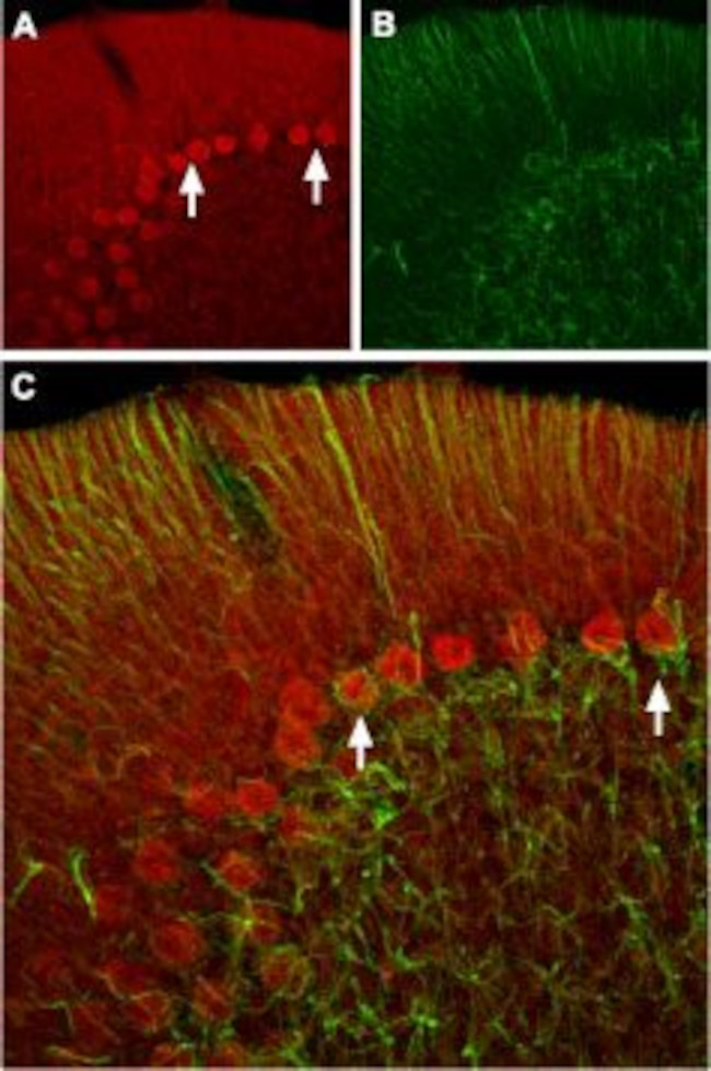
Expression of HCN2 in rat cerebellum - Immunohistochemical staining of rat cerebellum frozen sections using Anti-HCN2 Antibody (#APC-030 ). A. HCN2 (red) appears in Purkinje cells (arrows). B. Staining of astrocytes with mouse Anti-glial fibrillary acidic protein (GFAP, green demonstrates the restriction of HCN2 to neuronal cell bodies. C. Confocal merge of HCN2 and GFAP images demonstrates the respective localization of these proteins.
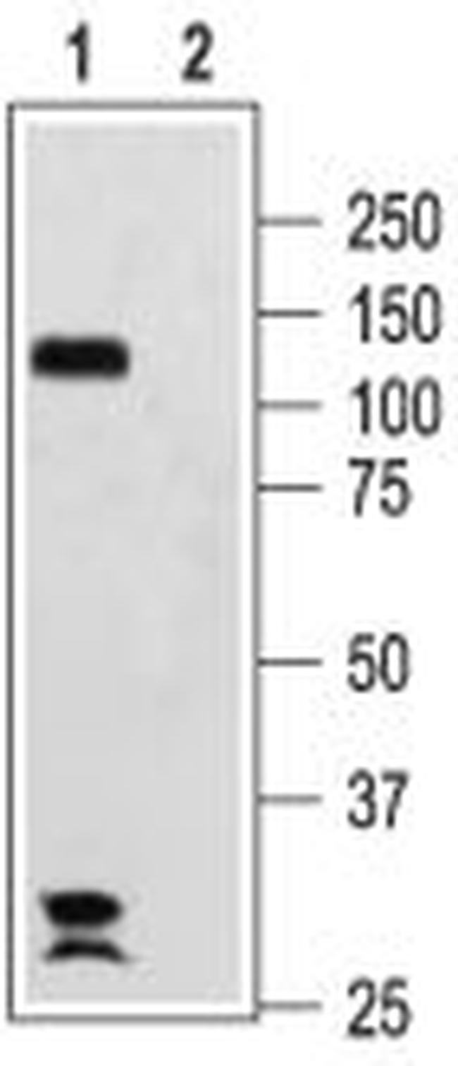
Western blotanalysis of rat brain membranes: - 1. Anti-HCN2 Antibody (#APC-030), (1:200).2. Anti-HCN2 Antibody , preincubated with HCN2 Blocking Peptide (#BLP-PC030).