


 全部商品分类


Anti-mouse IgG (H+L), F(ab‘) 2 Fragment (Alexa Fluor ® 488 Conjugate)

 下载产品说明书 下载COA 下载SDS
 用小程序,查商品更便捷


 收藏
收藏
 对比
对比 咨询
咨询
Anti-Mouse IgG (H+L) F(ab')2 Fragment antibody was conjugated to Alexa Fluor® 488 fluorescent dye under optimal conditions and formulated at 2 mg/ml. This F(ab')2 fragment product results in less non-specific binding, as it lacks the Fc domain that can bind to the cells with Fc receptors.
Product Usage Information
The optimal dilution of the anti-species antibody should be determined for each primary antibody by titration. However, a final dilution of 1:500 – 1:2000 should yield acceptable results for immunofluorescent and flow cytometry assays.
Specificity/Sensitivity
Species Reactivity:
Mouse
Supplied in 0.1 M sodium phosphate, 0.1 M sodium chloride, pH 7.5, 5 mM sodium azide. Store at 4°C. Do not aliquot the antibody. Protect from light. Do not freeze.
参考图片
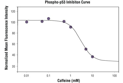
High content analysis of A439 cells exposed to varying concentrations of caffeine for 30 min prior to and 1.5 hr following a 100 mJ UV-treatment. With increasing concentrations of caffeine, a significant decrease (~2.5 fold) in phospho-p53 signal as compared to the UV-treated control was observed. When using phospho-p53 as a measurement, the IC50 of this compound was 2.95 mM. Data was generated on the Acumen® HCS platform using Anti-Mouse IgG (H+L), F(ab')2 Fragment (Alexa Fluor® 488 Conjugate).
Confocal immunofluorescent analysis of mouse cerebellum using CaMKII-α (6G9) Mouse mAb #50049 detected with Anti-mouse IgG (H+L), F(ab')2 Fragment (Alexa Fluor® 488 Conjugate) #4408 (green) and GFAP (D1F4Q) XP® Rabbit mAb #12389 detected with Anti-rabbit IgG (H+L), F(ab')2 Fragment (Alexa Fluor® 594 Conjugate) #8889 (red). Sections were mounted in ProLong® Gold Antifade Reagent with DAPI #8961 (blue).
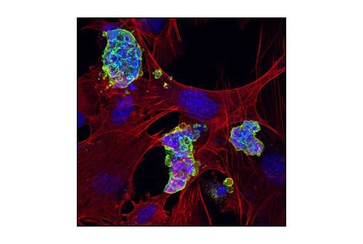
Confocal immunofluorescent analysis of mouse embryonic stem cells growing on mouse embryonic fibroblast (MEF) feeder cells using SSEA1 (MC480) Mouse mAb #4744 detected with anti-Mouse IgG (H+L), F(ab')2 Fragment (Alexa Fluor® 488 Conjugate) (green). Actin filaments have been labeled with DY-554 phalloidin (red). Blue pseudocolor = DRAQ5® #4084 (fluorescent DNA dye).
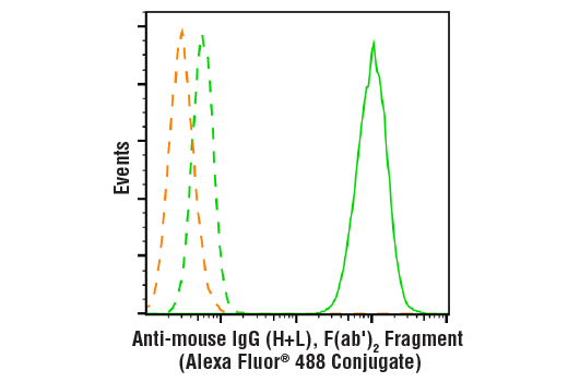
Flow cytometric analysis of HeLa cells using β-Actin (8H10D10) Mouse mAb #3700 (green solid line) compared to secondary antibody alone (green dashed line) or unstained cells (orange dashed line). Anti-mouse IgG (H+L), F(ab')2 Fragment (Alexa Fluor® 488 Conjugate) was used as a secondary antibody.