


 全部商品分类


Anti-rabbit IgG (H+L), F(ab‘) 2 Fragment (Alexa Fluor ® 647 Conjugate)

 下载产品说明书 下载COA 下载SDS
 用小程序,查商品更便捷


 收藏
收藏
 对比
对比 咨询
咨询
Anti-rabbit IgG (H+L) F(ab')2 Fragment was conjugated to Alexa Fluor® 647 fluorescent dye under optimal conditions and formulated at 2 mg/ml. This F(ab')2 fragment product results in less non-specific binding, as it lacks the Fc domain that can bind to the cells with Fc receptors.
Product Usage Information
The optimal dilution of the anti-species antibody should be determined for each primary antibody by titration. However, a final dilution of 1:500 – 1:2000 should yield acceptable results for immunofluorescent and flow cytometry assays.
Specificity/Sensitivity
Species Reactivity:
Rabbit
Supplied in 0.1 M sodium phosphate, 0.1 M sodium chloride, pH 7.5, 5 mM sodium azide. Store at 4°C. Do not aliquot the antibody. Protect from light. Do not freeze.
参考图片
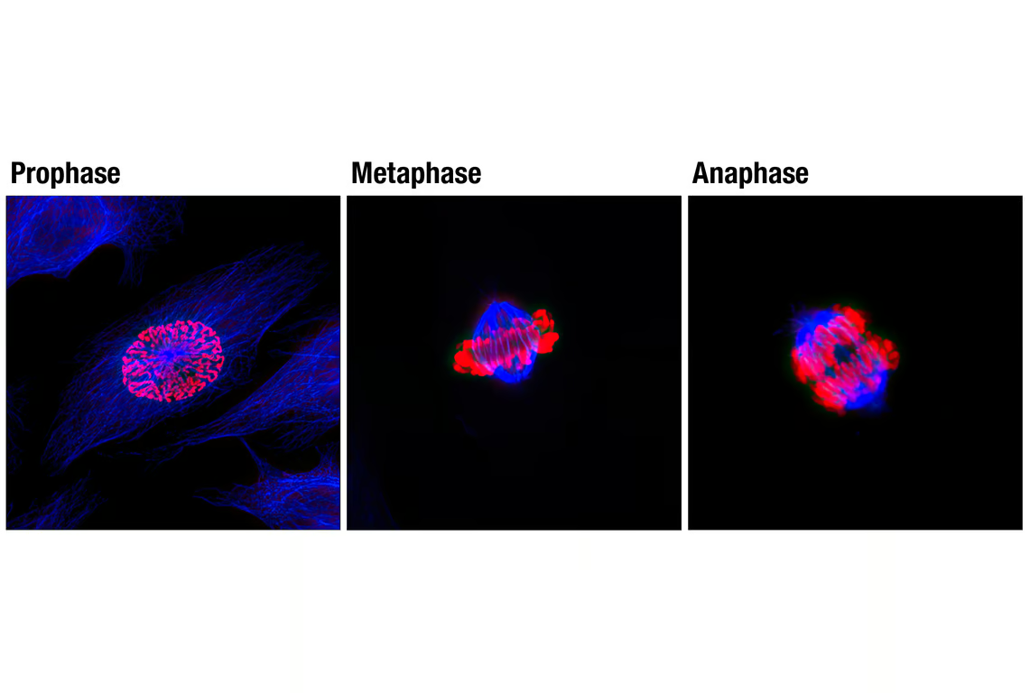
Confocal immunofluorescent analysis of mitotic HeLa cells using β-Tubulin (9F3) Rabbit mAb #2128 detected with Anti-rabbit IgG (H+L), F(ab')2 Fragment (Alexa Fluor® 647 Conjugate) (blue) and Phospho-Histone H3 (Ser10) (D2C8) XP® Rabbit mAb (Alexa Fluor® 555 Conjugate) #3475 (red).
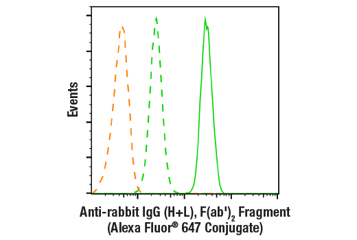
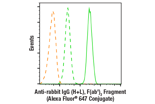
Flow cytometric analysis of Jurkat cells using Akt (pan) (C67E7) Rabbit mAb (Alexa Fluor® 647 Conjugate) #5186 (green solid line) compared to secondary antibody alone (green dashed line) or unstained cells (orange dashed line). Anti-rabbit IgG (H+L), F(ab')2 Fragment (Alexa Fluor® 647 Conjugate) was used as a secondary antibody.
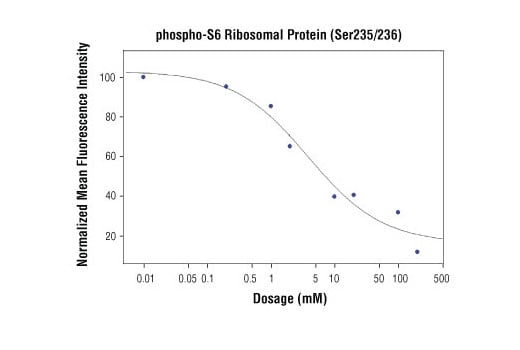
High content analysis of A549 cells exposed to varying concentrations of LY294002 (#9901) for 3 hrs, followed by 100 ng/mL EGF for 20 minutes. With increasing concentrations of LY294002, a significant decrease (~5 fold) in phospho-S6 Ribosomal Protein (Ser235/236) signal as compared to the uninhibited control was observed. When using phospho-S6 as a measurement, the IC50 of this compound was 3.06 μM. Data were generated on the Acumen® HCS platform using Anti-rabbit IgG (H+L), F(ab')2 Fragment (Alexa Fluor® 647 Conjugate).