


 全部商品分类


beta-Actin (13E5) Rabbit mAb


 下载产品说明书 下载COA 下载SDS
 用小程序,查商品更便捷


 收藏
收藏
 对比
对比 咨询
咨询
Monoclonal antibody is produced by immunizing animals with a synthetic peptide corresponding to residues near the amino-terminus of human β-actin protein.
Product Usage Information
| Application | Dilution |
|---|---|
| Western Blotting | 1:1000 |
| Simple Western™ | 1:10 - 1:50 |
| Immunohistochemistry (Paraffin) | 1:50 - 1:200 |
| Immunofluorescence (Frozen) | 1:100 - 1:400 |
| Immunofluorescence (Immunocytochemistry) | 1:100 - 1:400 |
| Flow Cytometry (Fixed/Permeabilized) | 1:100 - 1:400 |
Specificity/Sensitivity
Species Reactivity:
Human, Mouse, Rat, Monkey, Bovine, Pig
Supplied in 10 mM sodium HEPES (pH 7.5), 150 mM NaCl, 100 µg/ml BSA, 50% glycerol and less than 0.02% sodium azide. Store at –20°C. Do not aliquot the antibody.
For a carrier free (BSA and azide free) version of this product see product #93473.
参考图片
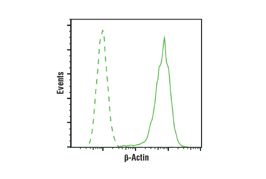
Flow cytometric analysis of HeLa cells using β-Actin (13E5) Rabbit mAb (solid line) compared to concentration-matched Rabbit (DA1E) mAb IgG XP® Isotype Control #3900 (dashed line). Anti-rabbit IgG (H+L), F(ab')2 Fragment (Alexa Fluor® 488 Conjugate) #4412 was used as a secondary antibody.
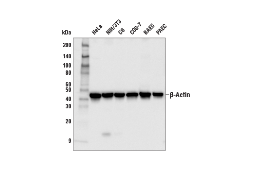
Western blot analysis of cell extracts from various cell lines using beta-Actin (13E5) Rabbit mAb.
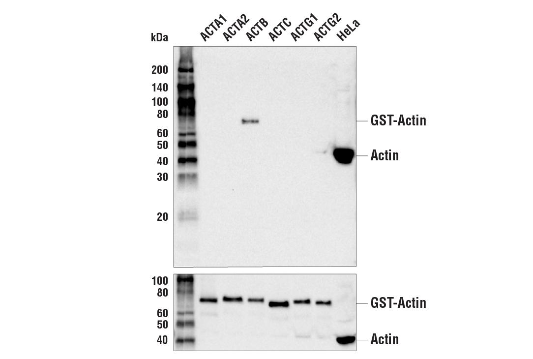
Western blot analysis of recombinant Actin isoforms using β-Actin (13E5) Rabbit mAb (upper) and Pan-Actin Antibody #4968 (lower).
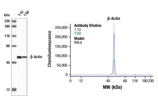
Simple Western™ analysis of lysates (0.1 mg/mL) from HeLa cells using ß-Actin (13E5) Rabbit mAb #4970. The virtual lane view (left) shows the target band (as indicated) at 1:10 and 1:50 dilutions of primary antibody. The corresponding electropherogram view (right) plots chemiluminescence by molecular weight along the capillary at 1:10 (blue line) and 1:50 (green line) dilutions of primary antibody. This experiment was performed under reducing conditions on the Jess™ Simple Western instrument from ProteinSimple, a BioTechne brand, using the 12-230 kDa separation module.
Immunohistochemical analysis of paraffin-embedded human heart using beta-Actin (13E5) Rabbit mAb. Note the lack of staining of cardiac actin.
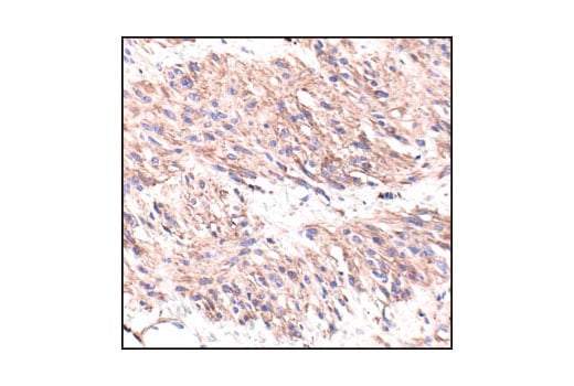
Immunohistochemical analysis of paraffin-embedded human leiomyoma using beta-Actin (13E5) Rabbit mAb.
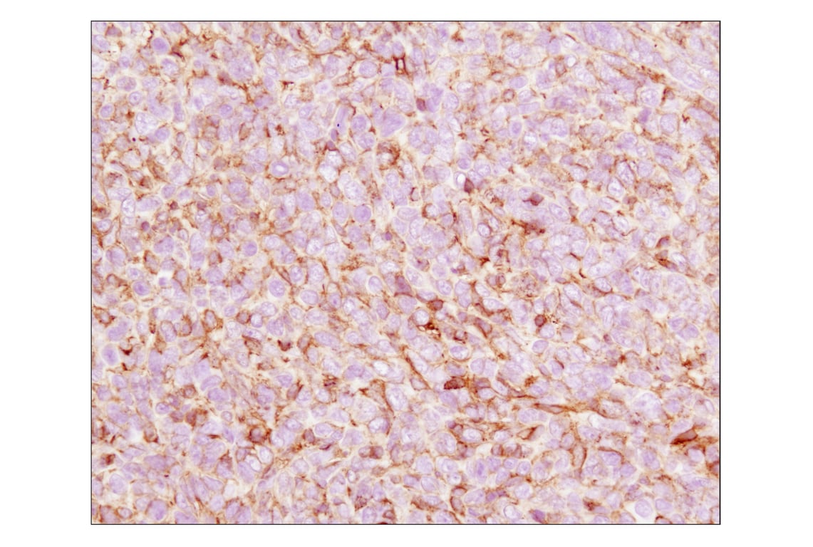
Immunohistochemical analysis of paraffin-embedded 4T1 syngeneic mouse tumor using β-actin (13E5) Rabbit mAb.
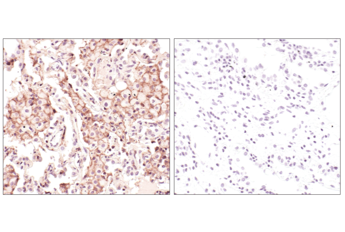
Immunohistochemical analysis of paraffin-embedded human lung carcinoma using beta-Actin (13E5) Rabbit mAb in the presence of control peptide (left) or beta-Actin Blocking Peptide #1025 (right).
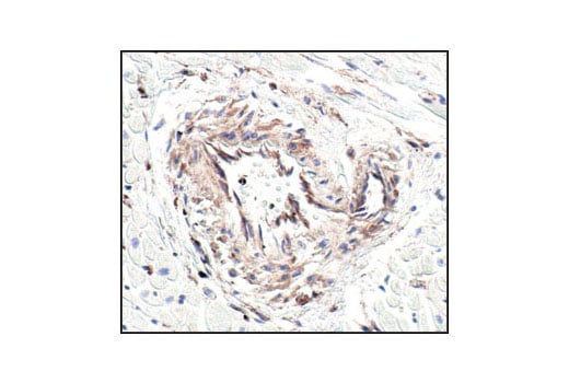
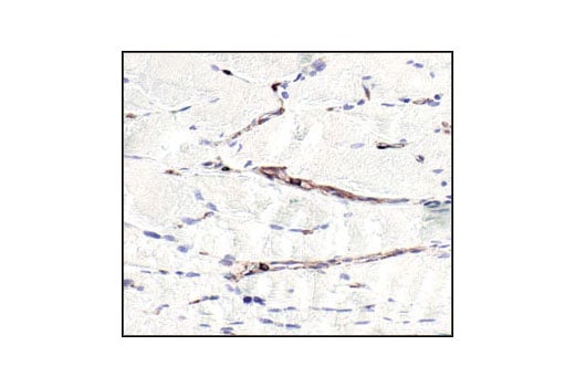
Immunohistochemical analysis of paraffin-embedded human skeletal muscle using beta-Actin (13E5) Rabbit mAb. Note the lack of staining of skeletal muscle actin.
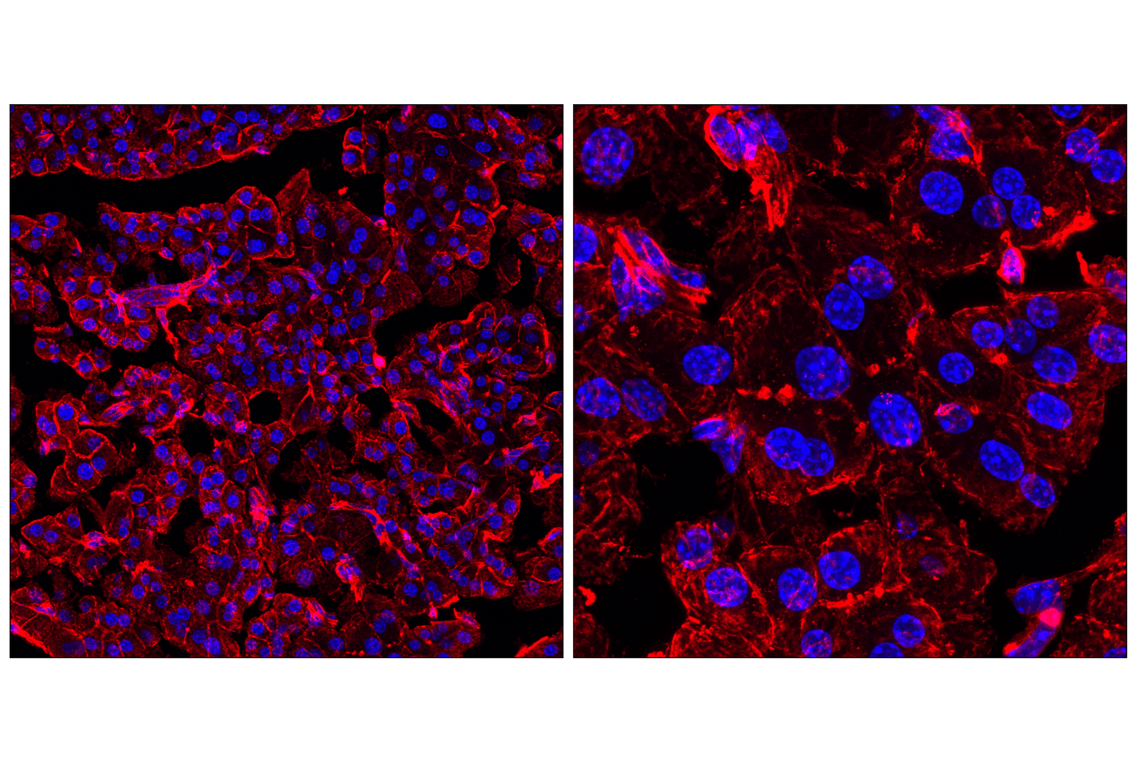
Confocal immunofluorescent analysis of fixed, frozen mouse pancreas at low magnification (left) and high magnification (right) using β-Actin (13E5) Rabbit mAb #4970 (red) and DAPI #4083 (blue).
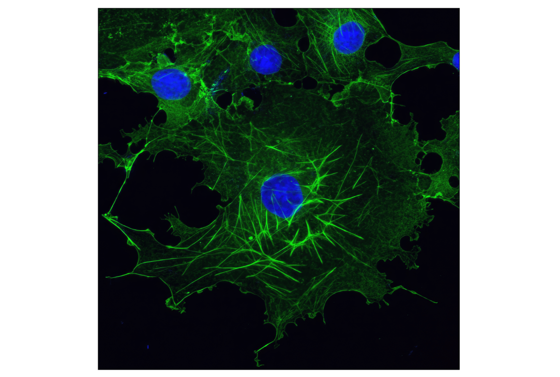
Confocal immunofluorescent analysis of COS-7 cells using β-Actin (13E5) Rabbit mAb (green). Blue pseudocolor = DRAQ5® #4084 (fluorescent DNA dye).