 全部商品分类


This antibody is prepared from goat antibodies and purified by immunoaffinity chromatography using antigen coupled to agarose beads.


Anti-rabbit IgG (H+L) was conjugated to DyLight 800 4X PEG fluorescent dye under optimal conditions and formulated at 1 mg/ml. Excitation is 777 nm and peak fluorescence emission is 794 nm.

Product Usage Information
The optimal dilution of the anti-species antibody should be determined by the user. However, the final dilutions below should yield acceptable results for the respective applications.Fluorescent western blotting: 1:30000In-Cell Western: 1:1000


Specificity/Sensitivity
Species Reactivity:
Rabbit


Proprietary buffer. Store at 4°C. Protect from Light. Do not freeze.
参考图片
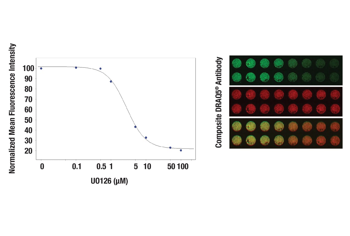
In-Cell Western™ analysis of A549 cells exposed to varying concentrations of U0126 (MEK1/2 Inhibitor) #9903 for 3 hours, followed by TPA (Phorbol-12-Myristate-13-Acetate) #9905 stimulation for 30 minutes. With increasing concentrations of U0126, a significant decrease (~5 fold) in Phospho p44/42 MAPK (Erk1/2) (Thr202/Tyr204) (D13.14.4E) XP® Rabbit mAb #4370 signal as compared to the TPA-stimulated control was observed. When using phospho-Erk as a measurement, the IC50 of this compound was 2.8 μM. Data and images were generated on the LI-COR Biosciences Odyssey Infrared Imaging System using Anti-rabbit IgG (H+L) (DyLight 800 4X PEG Conjugate). DRAQ5® #4084 (fluorescent DNA dye - red) is used for normalization.
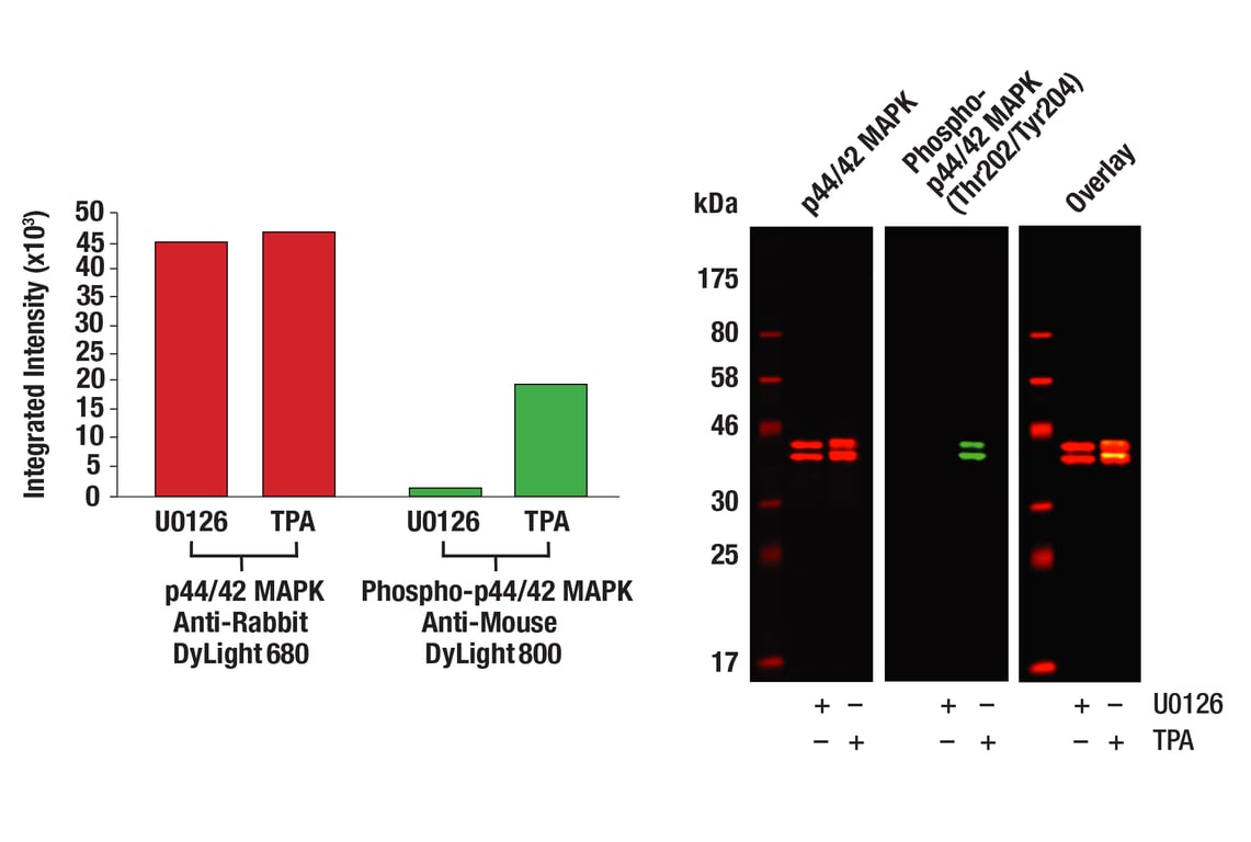
Western blot analysis of Jurkat cell lysates (#9194) treated with either U0126 (MEK 1/2 inhibitor) #9903 or TPA (12-O-Tetradecanoylphorbol-13-Acetate) #4174, using Phospho-p44/42 MAPK (Erk1/2) (Thr202/204) (D13.14.4E) XP® Rabbit mAb #4370 detected with Anti-rabbit IgG (H+L) (DyLight 800 4X PEG Conjugate) (green) and p44/42 MAPK (Erk1/2) (3A7) Mouse mAb #9107 detected with Anti-mouse IgG (H+L) (DyLight 680 Conjugate) #5470 (red). The array image pixel intensities obtained using a LI-COR Biosciences Odyssey Infrared Imaging System are shown in the panel to the left while corresponding fluorescent western blots are shown in the panel to the right.


 用小程序,查商品更便捷


