


 全部商品分类


Mouse (E7Q5L) mAb IgG2b Isotype Control

 下载产品说明书 下载COA 下载SDS
 用小程序,查商品更便捷


 收藏
收藏
 对比
对比 咨询
咨询
Product Usage Information
Important! This control antibody must be diluted to the same concentration (not dilution) as the specific antibody used in analysis. Higher background fluorescence may result if excessive amounts of isotype control are used. For protocol details, please reference the product page for the specific antibody used in analysis.
Specificity/Sensitivity
Supplied in 10 mM sodium HEPES (pH 7.5), 150 mM NaCl, 100 µg/ml BSA, 50% glycerol and less than 0.02% sodium azide. Store at –20°C. Do not aliquot the antibody.
参考图片
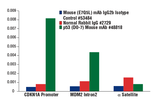
Chromatin immunoprecipitations were performed with cross-linked chromatin from 4 x 10⁶ HCT116 cells treated with UV (1000 J/m2 followed by a 3 hour recovery) and either Mouse (E7Q5L) mAb IgG2b Isotype Control, Normal Rabbit IgG #2729, or p53 (DO-7) Mouse mAb #48818 using SimpleChIP® Plus Enzymatic Chromatin IP Kit (Magnetic Beads) #9005. The enriched DNA was quantified by real-time PCR using SimpleChIP® Human CDKN1A Promoter Primers #6449, SimpleChIP® Human MDM2 Intron 2 Primers #90678, and SimpleChIP® Human α Satellite Repeat Primers #4486. The amount of immunoprecipitated DNA in each sample is represented as signal relative to the total amount of input chromatin, which is equivalent to one.
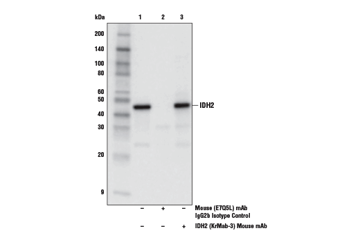
Immunoprecipitation of IDH2 from Jurkat cell extracts. Lane 1 is 10% input, lane 2 is Mouse (E7Q5L) mAb IgG2b Isotype Control, and lane 3 is IDH2 (KrMab-3) Mouse mAb #60322. Western blot analysis was performed using IDH2 (D8E3B) Rabbit mAb #56439. Anti-rabbit IgG, HRP-linked Antibody #7074 was used as the secondary antibody.
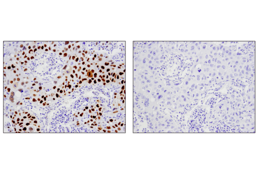
Immunohistochemical analysis of paraffin-embedded human lung carcinoma using p53 (DO-7) Mouse mAb #48818 (left) compared to concentration matched Mouse (E7Q5L) mAb IgG2b Isotype Control (right).
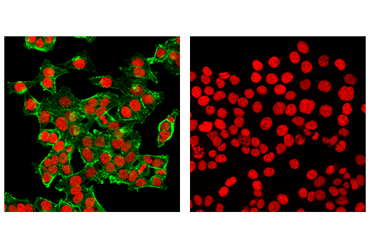
Confocal immunofluorescent analysis of HCT 116 cells using β-Actin (8H10D10) Mouse mAb #3700 (left, green) compared to concentration matched Mouse (E7Q5L) mAb IgG2b Isotype Control (right, green). Red = Propidium Iodide (PI)/RNase Staining Solution #4087.
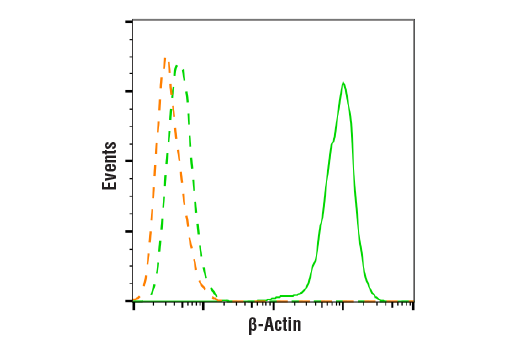
Flow cytometric analysis of HeLa cells using β-Actin (8H10D10) Mouse mAb #3700 (green solid line) compared to concentration-matched Mouse (E7Q5L) mAb IgG2b Isotype Control (green dashed line) or secondary antibody alone (orange dashed line). Anti-mouse IgG (H+L), F(ab')2 Fragment (Alexa Fluor® 488 Conjugate) #4408 was used as a secondary antibody.