


 全部商品分类


 用小程序,查商品更便捷


Product Usage Information
Important! This control antibody must be diluted to the same concentration (not dilution) as the specific antibody used in analysis. Higher background fluorescence may result if excessive amounts of isotype control are used. For protocol details, please reference the product page for the specific antibody used in analysis.
Specificity/Sensitivity
Supplied in 10 mM sodium HEPES (pH 7.5), 150 mM NaCl, 100 µg/ml BSA, 50% glycerol and less than 0.02% sodium azide. Store at –20°C. Do not aliquot the antibody.
For a carrier free (BSA and azide free) version of this product see product #87582.
参考图片
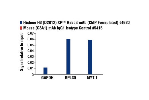
Chromatin immunoprecipitations were performed using digested chromatin from HeLa cells and the indicated antibodies. Purified DNA was analyzed by quantitative real-time PCR, using SimpleChIP® Human GAPDH Exon 1 Primers #5516, SimpleChIP® Human RPL30 Exon 3 Primers #7014, and SimpleChIP® Human MYT-1 Exon 1 Primers #4493. The relative abundance of each DNA sequence enriched by Mouse (G3A1) mAb IgG1 Isotype Control (red) is compared to the amount of the same DNA sequence enriched by the histone H3-specific immunoprecipitations (blue).
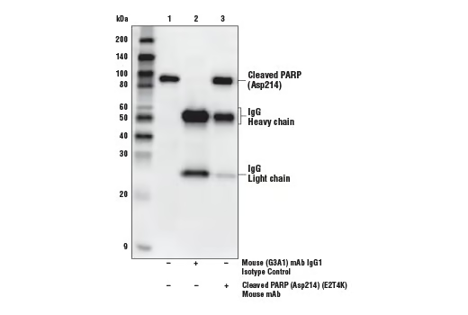
Immunoprecipitation of cleaved PARP (Asp214) from serum-starved HeLa cells treated with Staurosporine #9953 (1 μM, 3 hr). Lane 1 is 10% input, lane 2 is Mouse (G3A1) mAb IgG1 Isotype Control, and lane 3 is Cleaved PARP (Asp214) (E2T4K) Mouse mAb #32563. Western blot was performed using Cleaved PARP (Asp214) (E2T4K) Mouse mAb #32563. Anti-mouse IgG, HRP-linked Antibody #7076 was used as a secondary antibody.
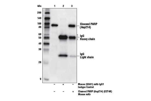
Immunoprecipitation of cleaved PARP (Asp214) from serum-starved HeLa cells treated with Staurosporine #9953 (1 μM, 3 hr). Lane 1 is 10% input, lane 2 is Mouse (G3A1) mAb IgG1 Isotype Control, and lane 3 is Cleaved PARP (Asp214) (E2T4K) Mouse mAb #32563. Western blot was performed using Cleaved PARP (Asp214) (E2T4K) Mouse mAb #32563. Anti-mouse IgG, HRP-linked Antibody #7076 was used as a secondary antibody.
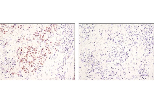
Immunohistochemical analysis of paraffin-embedded human seminoma using Nanog (1E6C4) Mouse mAb #4893 (left) or Mouse (G3A1) mAb IgG1 Isotype Control (right).
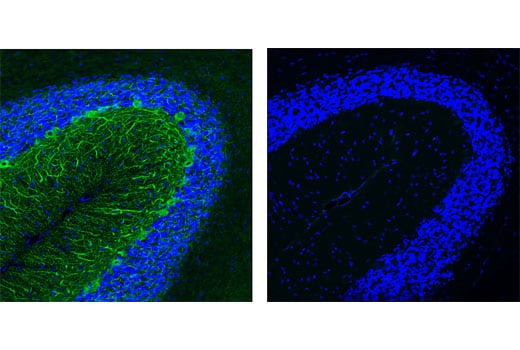
Confocal immunofluorescent analysis of normal rat cerebellum using Neurofilament H (RMdO 20) Mouse mAb #2836 (green, left) compared to concentration matched Mouse (G3A1) mAb IgG1 Isotype Control (green, right). Blue pseudocolor = DRAQ5® #4084 (fluorescent DNA dye).
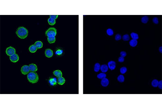
Confocal immunofluorescent analysis of HT-29 cells using α-Tubulin (DM1A) Mouse mAb #3873 (green, left) compared to concentration matched Mouse (G3A1) mAb IgG1 Isotype Control (green, right). Blue pseudocolor = DRAQ5® #4084 (fluorescent DNA dye).
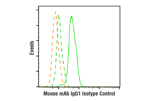
Flow cytometric analysis of A431 cells using Pan-Keratin (C11) Mouse mAb #4545 (green solid line) compared to concentration-matched Mouse (G3A1) mAb IgG1 Isotype Control (green dashed line) or secondary antibody alone (orange dashed line). Anti-mouse IgG (H+L), F(ab')2 Fragment (Alexa Fluor® 488 Conjugate) #4408 was used as a secondary antibody.