


 全部商品分类


Anti-mouse IgG (H+L) (DyLight™ 680 Conjugate)

 下载产品说明书 下载COA 下载SDS
 用小程序,查商品更便捷


 收藏
收藏
 对比
对比 咨询
咨询
This antibody is prepared from goat antibodies and purified by immunoaffinity chromatography using antigen coupled to agarose beads.
Anti-mouse IgG (H+L) was conjugated to DyLight 680 fluorescent dye under optimal conditions and formulated at 1 mg/ml. Excitation is 684 nm and peak fluorescence emission is 715 nm.
Product Usage Information
The optimal dilution of the anti-species antibody should be determined by the user. However, the final dilutions below should yield acceptable results for the respective applications.Fluorescent western blotting: 1:15000In-Cell Western: 1:1000
Specificity/Sensitivity
Species Reactivity:
Mouse
Supplied in 100 mM PBS, pH 7.2, containing 1% BSA and 0.02% sodium azide. Store at 4°C. Protect from Light. Do not freeze.
参考图片
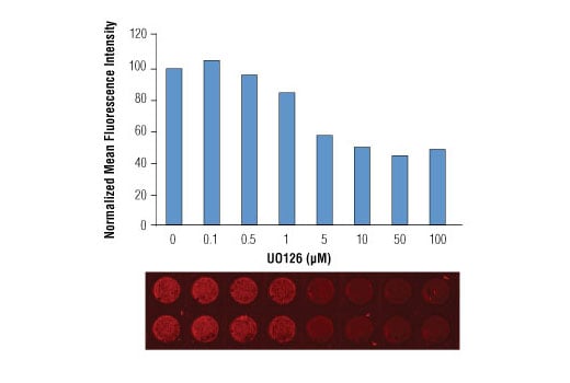
In-Cell Western™ analysis of A549 cells exposed to varying concentrations of U0126 (MEK1/2 Inhibitor) #9903 for 3 hours, followed by TPA (Phorbol-12-Myristate-13-Acetate) #9905 stimulation for 30 minutes. With increasing concentrations of U0126, a significant decrease (~2.5 fold) in Phospho-p44/42 MAPK (Erk1/2) (Thr202/Tyr204) (E10) Mouse mAb #9106 signal as compared to the TPA-stimulated control was observed. Data and images were generated on the LI-COR® Biosciences Odyssey® Infrared Imaging System using Anti-mouse IgG (H+L) (DyLight™ 680 Conjugate).
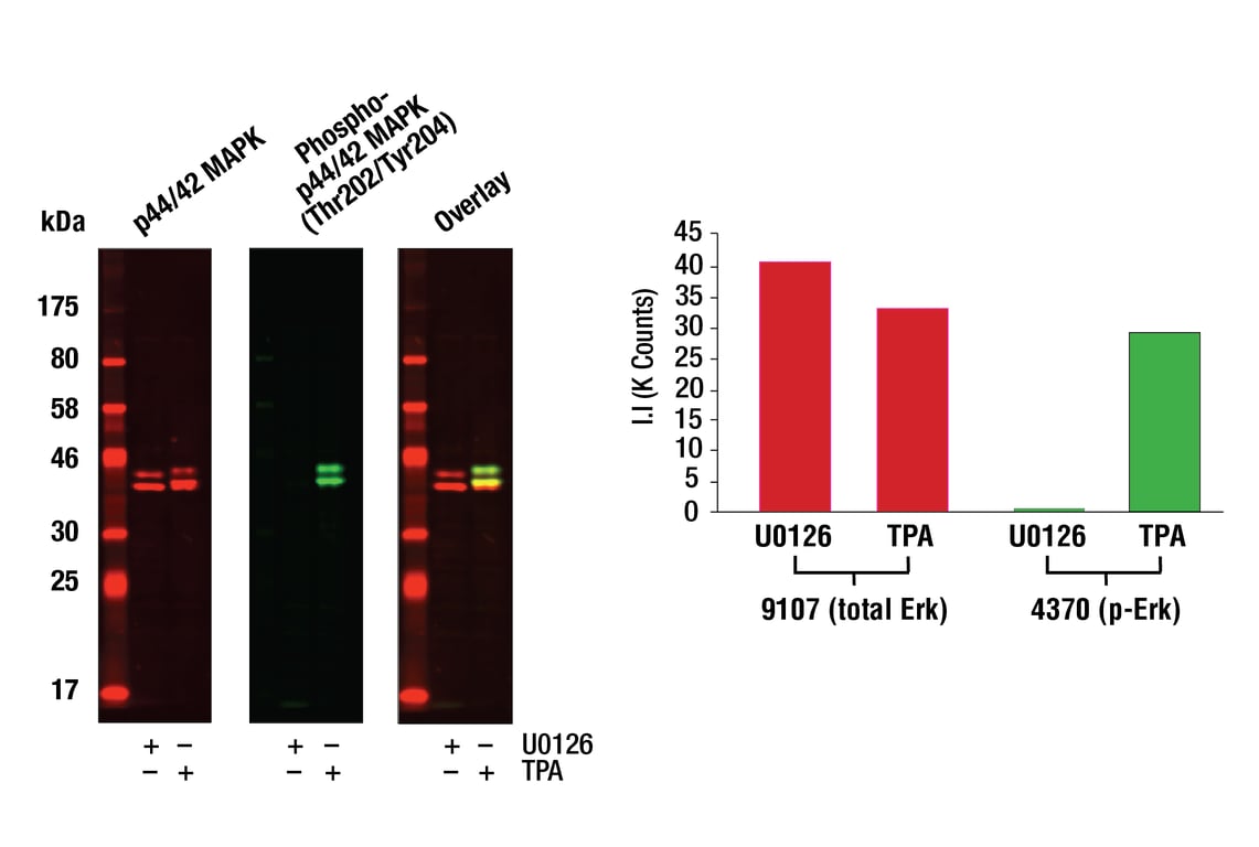
Western blot analysis of Jurkat cell lysates (#9194) treated with either U0126 (MEK 1/2 inhibitor) #9903 or TPA (12-O-Tetradecanoylphorbol-13-Acetate) #4174, using Phospho-p44/42 MAPK (Erk1/2) (Thr202/204) (D13.14.4E) XP® Rabbit mAb #4370 detected with Anti-rabbit IgG (H+L) (DyLight 800 Conjugate) #5151 (green) and p44/42 MAPK (Erk1/2) (3A7) Mouse mAb #9107 detected with Anti-mouse IgG (H+L) (DyLight 680 Conjugate) (red). The array image pixel intensities obtained using a LI-COR Biosciences Odyssey Infrared Imaging System are shown in the top figure while corresponding fluorescent western blots are shown in the bottom figure.