


 全部商品分类


PD-1 (Intracellular Domain) (D4W2J) XP ® Rabbit mAb

 下载产品说明书 下载COA 下载SDS
 用小程序,查商品更便捷


 收藏
收藏
 对比
对比 咨询
咨询
Monoclonal antibody is produced by immunizing animals with a synthetic peptide corresponding to residues surrounding Ala274 of human PD-1 protein.
Product Usage Information
| Application | Dilution |
|---|---|
| Western Blotting | 1:1000 |
| Immunoprecipitation | 1:200 |
| IHC Leica Bond | 1:100 - 1:400 |
| Immunohistochemistry (Paraffin) | 1:100 - 1:400 |
| Immunofluorescence (Immunocytochemistry) | 1:200 - 1:400 |
| Flow Cytometry (Fixed/Permeabilized) | 1:100 - 1:200 |
Specificity/Sensitivity
Species Reactivity:
Human
Supplied in 10 mM sodium HEPES (pH 7.5), 150 mM NaCl, 100 µg/ml BSA, 50% glycerol and less than 0.02% sodium azide. Store at –20°C. Do not aliquot the antibody.
For a carrier free (BSA and azide free) version of this product see product #63815.
参考图片
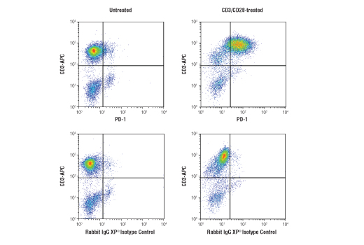
Flow cytometric analysis of fixed and permeabilized human peripheral blood mononuclear cells, untreated (left column) or treated with anti-CD3 (10ug/ml, 72hr) and anti-CD28 (5ug/ml, 72 hr; right column), using PD-1 (Intracellular Domain) (D4W2J) XP® Rabbit mAb #86163 (top row) or concentration-matched Rabbit (DA1E) mAb IgG XP® Isotype Control #3900 (bottom row), and co-stained with CD3 (UCHT1) Mouse mAb (APC Conjugate) #19881. Anti-rabbit IgG (H+L), F(ab')2 Fragment (Alexa Fluor® 488 Conjugate) #4412 was used as a secondary antibody.
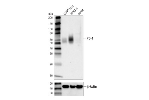
Western blot analysis of extracts from human CD4+ T cells, MOLT-4, and Jurkat cells using PD-1 (Intracellular Domain) (D4W2J) XP® Rabbit mAb (upper), and β-Actin (D6A8) Rabbit mAb #8457 (lower). CD4+ T cells were purified from human blood and stimulated for 9 days using beads coated with CD3 and CD28 antibodies in the presence of human interleukin-2 (hIL-2) #8907 (6.7 ng/ml).
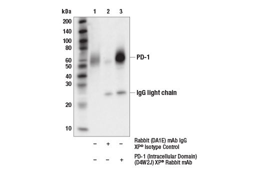
Immunoprecipitation of PD-1 protein from Molt-4 cell extracts. Lane 1 is 10% input, lane 2 is Rabbit (DA1E) mAb IgG XP® Isotype Control #3900, and lane 3 is PD-1 (Intracellular Domain) (D4W2J) XP® Rabbit mAb. Western blot analysis was performed using PD-1 (Intracellular Domain) (D4W2J) XP® Rabbit mAb.
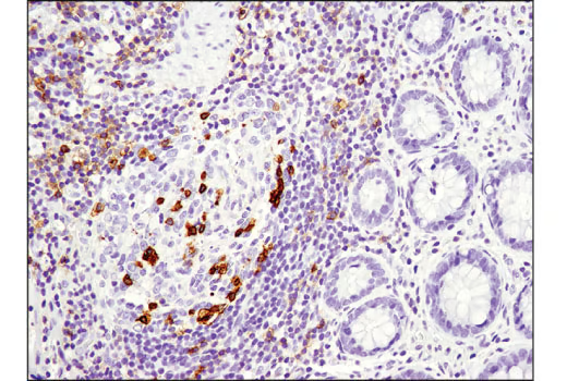
Immunohistochemical analysis of paraffin-embedded human infiltrating papillary carcinoma of the breast using PD-1 (Intracellular Domain) (D4W2J) XP® Rabbit mAb performed on the Leica® BOND™ Rx.
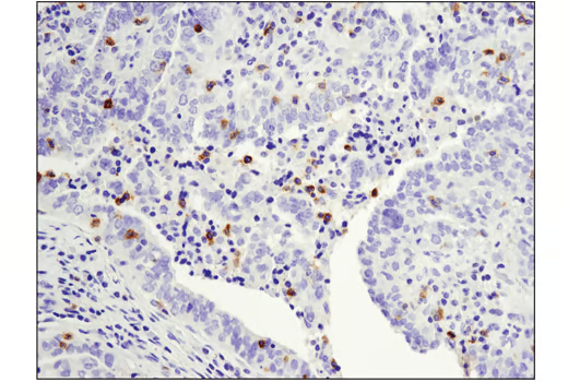
Immunohistochemical analysis of paraffin-embedded human colon carcinoma using PD-1 (Intracellular Domain) (D4W2J) XP® Rabbit mAb.
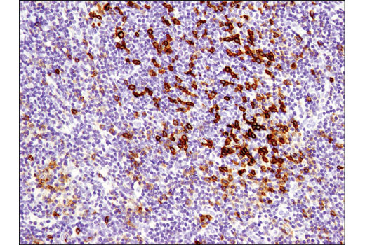
Immunohistochemical analysis of paraffin-embedded human B-cell non-Hodgkin lymphoma using PD-1 (Intracellular Domain) (D4W2J) XP® Rabbit mAb.
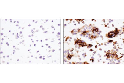
Immunohistochemical analysis of paraffin-embedded 293 cell pellets, control (left) or PD-1 transfected (right), using PD-1 (Intracellular Domain) (D4W2J) XP® Rabbit mAb.
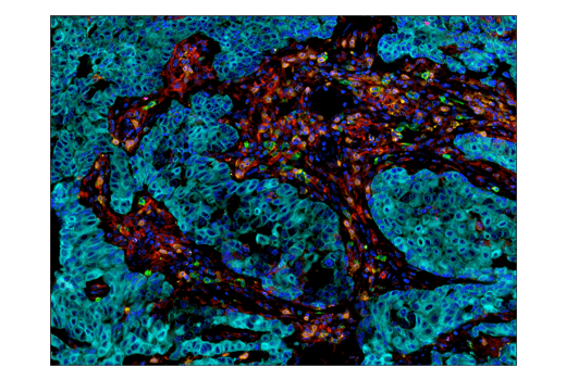
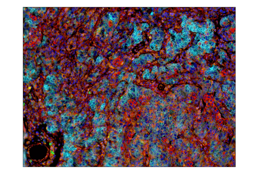
Multiplex immunohistochemical analysis of paraffin-embedded human breast carcinoma usng PD-1 (Intracellular Domain) (D4W2J) XP® rabbit mAb (green), PD-L1 (E1L3N®) XP® rabbit mAb #13684 (red), LAG3 (D2G4O™) XP® rabbit mAb #15372 (magenta), TIM-3 (D5D5R™) XP® rabbit mAb #45208 (yellow), CD8α (C8/144B) mouse mAb #70306 (orange), and Pan-keratin (C11) mouse mAb #4545 (cyan).
Multiplex immunohistochemical analysis of paraffin-embedded human ovarian carcinoma using PD-1 (Intracellular Domain) (D4W2J) XP® rabbit mAb (green), B7-H3 (D9M2L) XP® rabbit mAb #14058 (red), B7-H4 (D1M8I) XP® rabbit mAb (cyan), LAG3 (D2G4O™) XP® rabbit mAb #15372 (magenta), TIM-3 (D5D5R™) XP® rabbit mAb #45208 (yellow), and VISTA (D1L2G™) XP® rabbit mAb #64953 (orange).
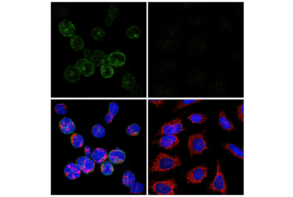
Confocal immunofluorescent analysis of MOLT-4 cells (left, positive) or HeLa cells (right, negative) using PD-1 (Intracellular Domain) (D4W2J) XP® Rabbit mAb (green), Cytochrome c (6H2.B4) Mouse mAb #12963 (red), and DAPI #4083 (blue).