


 全部商品分类
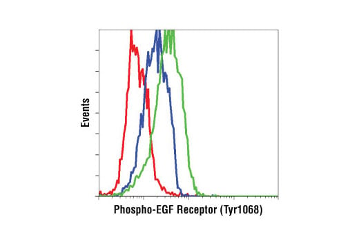


 用小程序,查商品更便捷


Monoclonal antibody is produced by immunizing animals with a synthetic phosphopeptide corresponding to residues surrounding Tyr1068 of human EGF receptor.
Product Usage Information
| Application | Dilution |
|---|---|
| Western Blotting | 1:1000 |
| Fluorescent Western | 1:1000 |
| Simple Western™ | 1:50 - 1:250 |
| Immunohistochemistry (Paraffin) | 1:200 - 1:800 |
| Immunofluorescence (Immunocytochemistry) | 1:400 - 1:1600 |
| Flow Cytometry (Fixed/Permeabilized) | 1:1600 |
Specificity/Sensitivity
Species Reactivity:
Human, Mouse, Rat, Monkey
Supplied in 10 mM sodium HEPES (pH 7.5), 150 mM NaCl, 100 µg/ml BSA, 50% glycerol and less than 0.02% sodium azide. Store at –20°C. Do not aliquot the antibody.
For a carrier-free (BSA and azide free) version of this product see product #48576.
参考图片
Flow cytometric analysis of A549 cells, untreated (blue) or EGF-treated (green), using Phospho-EGF Receptor (Tyr1068) (D7A5) XP® Rabbit mAb compared to concentration matched XP® Rabbit (DA1E) mAb IgG Isotype Control #3900 (red).
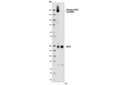
Western blot analysis of extracts of BxPC-3 cells, untreated or EGF-stimulated, using Phospho-EGF Receptor (Tyr1068) (D7A5) XP® Rabbit mAb (upper) and EGF Receptor Antibody #2232 (lower).
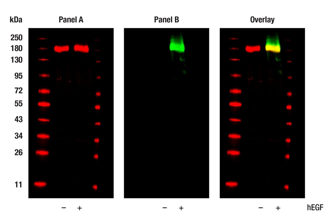
Western blot analysis of extracts from serum-starved A431 cells, untreated (-) or treated with hEGF (100 ng/ml, 5 min; +), using EGF Receptor (1F4) Mouse mAb #2239 (Panel A) and Phospho-EGF Receptor (Tyr1068) (D7A5) XP® Rabbit mAb #3777 (Panel B). Anti-rabbit IgG (H+L) (DyLight 800 4X PEG Conjugate) #5151 (green) and Anti-mouse IgG (H+L) (DyLight 680 Conjugate) #5470 (red) were used as secondary antibodies.
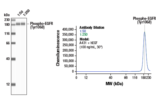
Simple Western™ analysis of lysates (0.1 mg/mL) from A431 cells treated with human EGF (100 ng/mL, 30”) using Phospho-EGF Receptor (Tyr1068) (D7A5) XP® Rabbit mAb #3777. The virtual lane view (left) shows a single target band (as indicated) at 1:50 and 1:250 dilutions of primary antibody. The corresponding electropherogram view (right) plots chemiluminescence by molecular weight along the capillary at 1:50 (blue line) and 1:250 (green line) dilutions of primary antibody. This experiment was performed under reducing conditions on the Jess™ Simple Western instrument from ProteinSimple, a BioTechne brand, using the 12-230 kDa separation module.
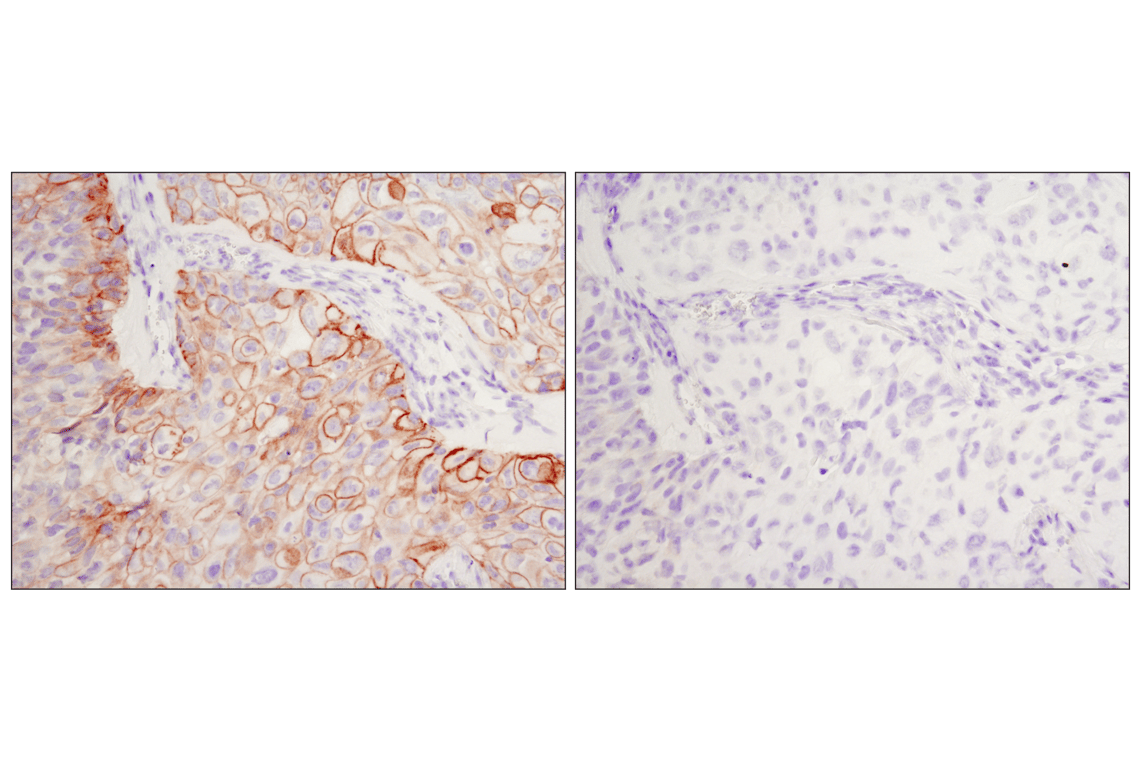
Immunohistochemical analysis of paraffin-embedded HCC827 xenograft, control (left) or λ phosphatase-treated (right), using Phospho-EGF Receptor (Tyr1068) (D7A5) XP® Rabbit mAb.
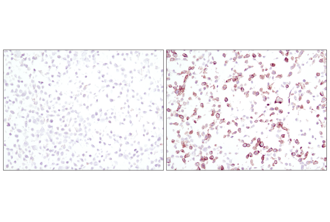
Immunohistochemical analysis using Phospho-EGF Receptor (Tyr1068) (D7A5) XP® Rabbit mAb on SignalSlide™ Phospho-EGF Receptor IHC Controls #8102 (paraffin-embedded KYSE450 cell pellets, untreated (left) or EGF-treated (right)).
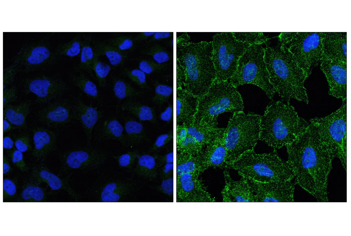
Confocal immunofluorescent analysis of HeLa cells, untreated (left) or EGF-treated (right), using Phospho-EGF Receptor (Tyr1068) (D7A5) XP® Rabbit mAb (green) and DRAQ5® #4084 (blue pseudocolor).