


 全部商品分类


BD Pharmingen™ Propidium Iodide Staining Solution


 下载产品说明书 下载SDS
 用小程序,查商品更便捷


 收藏
收藏
 对比
对比 咨询
咨询
参考图片
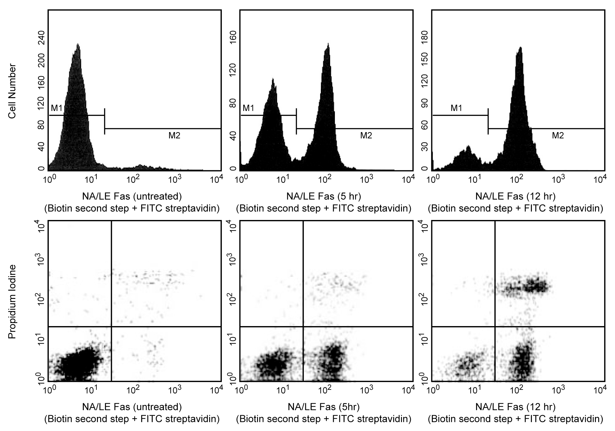
Flow cytometric analysis of cells following induction of apoptosis. Jurkat leukemia cells were left untreated (left top & left bottom panels) or treated for 5 hours (middle top & middle bottom panels) or 12 hours (right top & right bottom panels) with anti-human Fas antibody (clone DX2, Cat. No. 555670) and Protein G (the addition of Protein G enhances the ability of DX2 to induce apoptosis, presumably by cross-linking Fas). Cells were incubated with Annexin V-Biotin, (Cat. No. 556417) followed by incubation with SAv-FITC (Cat. No. 554060) in Propidium Iodide (PI) Staining Solution (Cat. No. 556463). Cells were then analyzed by flow cytometry. Untreated cells were primarily Annexin V-Biotin and PI negative, indicating that they were viable and not undergoing apoptosis. After a 5 hour treatment with DX2, there were two populations of cells: Cells undergoing apoptosis (Annexin V-Biotin positive and PI negative), and cells that were viable and not undergoing apoptosis (Annexin V-Biotin and PI negative). After a 12 hour treatment with DX2, three populations of cells were identified: Cells that had already died or were in late stage of apoptosis (Annexin V-Biotin and PI positive), cells undergoing apoptosis (Annexin V-Biotin positive and PI negative), and cells that were viable and not undergoing apoptosis (Annexin V-Biotin and PI negative).
Flow cytometric analysis of cells following induction of apoptosis. Jurkat leukemia cells were left untreated (left top & left bottom panels) or treated for 5 hours (middle top & middle bottom panels) or 12 hours (right top & right bottom panels) with anti-human Fas antibody (clone DX2, Cat. No. 555670) and Protein G (the addition of Protein G enhances the ability of DX2 to induce apoptosis, presumably by cross-linking Fas). Cells were incubated with Annexin V-Biotin, (Cat. No. 556417) followed by incubation with SAv-FITC (Cat. No. 554060) in Propidium Iodide (PI) Staining Solution (Cat. No. 556463). Cells were then analyzed by flow cytometry. Untreated cells were primarily Annexin V-Biotin and PI negative, indicating that they were viable and not undergoing apoptosis. After a 5 hour treatment with DX2, there were two populations of cells: Cells undergoing apoptosis (Annexin V-Biotin positive and PI negative), and cells that were viable and not undergoing apoptosis (Annexin V-Biotin and PI negative). After a 12 hour treatment with DX2, three populations of cells were identified: Cells that had already died or were in late stage of apoptosis (Annexin V-Biotin and PI positive), cells undergoing apoptosis (Annexin V-Biotin positive and PI negative), and cells that were viable and not undergoing apoptosis (Annexin V-Biotin and PI negative).