


 全部商品分类
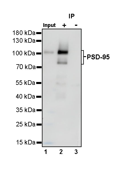


 用小程序,查商品更便捷


WB
1:1000IHC-P
1:500IP
1:50
PSD-95 is a member of the membrane-associated guanylate kinase (MAGUK) family. With PSD-93 it is recruited into the same NMDA receptor and potassium channel clusters. These two MAGUK proteins may interact at postsynaptic sites to form a multimeric scaffold for the clustering of receptors, ion channels, and associated signaling proteins. PSD-95 is the best studied member of the MAGUK-family of PDZ domain-containing proteins. Like all MAGUK-family proteins, its basic structure includes three PDZ domains, an SH3 domain, and a guanylate kinase-like domain (GK) connected by disordered linker regions. It is almost exclusively located in the post synaptic density of neurons, and is involved in anchoring synaptic proteins. Its direct and indirect binding partners include neuroligin, NMDA receptors, AMPA receptors, and potassium channels. It plays an important role in synaptic plasticity and the stabilization of synaptic changes during long-term potentiation.
12 months from date of receipt / reconstitution, -20 °C as supplied
参考图片
WB result of PSD-95 Rabbit mAb
Primary antibody: PSD-95 Rabbit mAb at 1/1000 dilution
Lane 1: mouse spleen lysate 20 µg
Lane 2: mouse brain lysate 20 µg
Negative control: mouse spleen lysate
Secondary antibody: Goat Anti-Rabbit IgG, (H+L), HRP conjugated at 1/10000 dilution
Predicted MW: 80 kDa
Observed MW: 80, 105 kDa
Exposure time: 120 s
WB result of PSD-95 Rabbit mAb
Primary antibody: PSD-95 Rabbit mAb at 1/1000 dilution
Lane 1: rat brain lysate 20 µg
Secondary antibody: Goat Anti-Rabbit IgG, (H+L), HRP conjugated at 1/10000 dilution
Predicted MW: 80 kDa
Observed MW: 80, 105 kDa
Exposure time: 120 s
IHC shows positive staining in paraffin-embedded human retina. Anti-PSD-95 antibody was used at 1/500 dilution, followed by a HRP Polymer for Mouse & Rabbit IgG (ready to use). Counterstained with hematoxylin. Heat mediated antigen retrieval with Tris/EDTA buffer pH9.0 was performed before commencing with IHC staining protocol.
IHC shows positive staining in paraffin-embedded mouse cerebral cortex. Anti-PSD-95 antibody was used at 1/500 dilution, followed by a HRP Polymer for Mouse & Rabbit IgG (ready to use). Counterstained with hematoxylin. Heat mediated antigen retrieval with Tris/EDTA buffer pH9.0 was performed before commencing with IHC staining protocol.
IHC shows positive staining in paraffin-embedded rat cerebral cortex. Anti-PSD-95 antibody was used at 1/500 dilution, followed by a HRP Polymer for Mouse & Rabbit IgG (ready to use). Counterstained with hematoxylin. Heat mediated antigen retrieval with Tris/EDTA buffer pH9.0 was performed before commencing with IHC staining protocol.
IHC shows positive staining in paraffin-embedded rat retina. Anti-PSD-95 antibody was used at 1/500 dilution, followed by a HRP Polymer for Mouse & Rabbit IgG (ready to use). Counterstained with hematoxylin. Heat mediated antigen retrieval with Tris/EDTA buffer pH9.0 was performed before commencing with IHC staining protocol.