


 全部商品分类
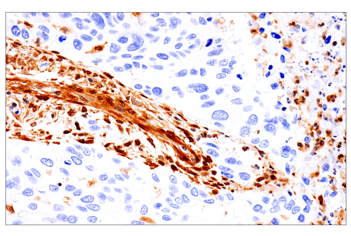


PTEN and PDK1 Antibody Sampler Kit II

 下载产品说明书 下载SDS
 用小程序,查商品更便捷


 收藏
收藏
 对比
对比 咨询
咨询
The PTEN and PDK1 Antibody Sampler Kit II provides an economical means to evaluate two key enzymes that regulate multiple signaling pathways. The kit includes enough antibodies to perform two western blot experiments with each primary antibody.
参考图片
Immunohistochemical analysis of paraffin-embedded human squamous cell lung carcinoma using PTEN (D4.3) XP® Rabbit mAb performed on the Leica BOND RX.
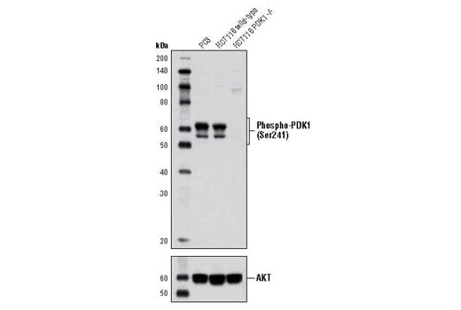
Western blot analysis of extracts from PC3 cells, HCT116 wild-type and HCT116 PDK1 -/- cells using Phospho-PDK1 (Ser241) (C49H2) Rabbit mAb (upper) and Akt (pan) (C67E7) Rabbit mAb #4691 (lower). (HCT116 wild-type and HCT116 PDK1 -/- cells were kindly provided by Dr. Bert Vogelstein, Johns Hopkins University, Baltimore, MD).
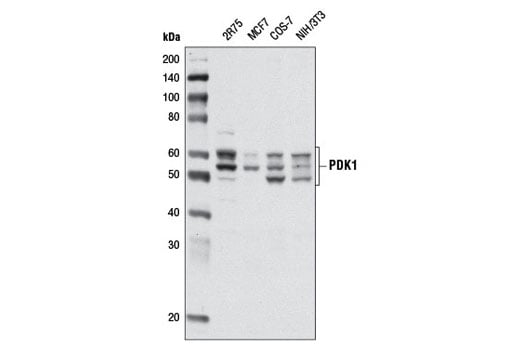
Western blot analysis of extracts from various cell lines using PDK1 (D37A7) Rabbit mAb.
After the primary antibody is bound to the target protein, a complex with HRP-linked secondary antibody is formed. The LumiGLO® is added and emits light during enzyme catalyzed decomposition.
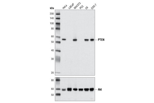
Western blot analysis of extracts from various cell lines using PTEN (D4.3) XP® Rabbit mAb (upper) and Akt (pan) (C67E7) Rabbit mAb #4691 (lower).
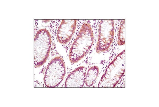
Immunohistochemical analysis of paraffin-embedded human colon using PTEN (D4.3) XP® Rabbit mAb.
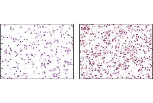
Immunohistochemical analysis using PTEN (D4.3) XP® Rabbit mAb on SignalSlide(TM) PTEN IHC Controls #8106 (paraffin-embedded LNCaP (left) and NIH/3T3 (right) cells).
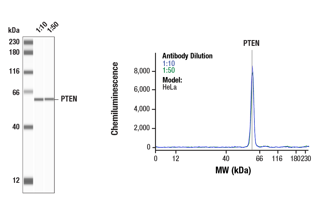
Simple Western™ analysis of lysates (1 mg/mL) from HeLa cells using PTEN (D4.3) XP® Rabbit mAb #9188. The virtual lane view (left) shows the target band (as indicated) at 1:10 and 1:50 dilutions of primary antibody. The corresponding electropherogram view (right) plots chemiluminescence by molecular weight along the capillary at 1:10 (blue line) and 1:50 (green line) dilutions of primary antibody. This experiment was performed under reducing conditions on the Jess™ Simple Western instrument from ProteinSimple, a BioTechne brand, using the 12-230 kDa separation module.
Immunohistochemical analysis of paraffin-embedded human esophageal adenocarcinoma using PTEN (D4.3) XP® Rabbit mAb performed on the Leica BOND RX.
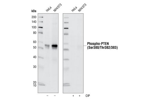
Western blot analysis of extracts from HeLa and NIH/3T3 cells, using Phospho-PTEN (Ser380/Thr382/383) (44A7) Rabbit mAb. Membranes were either left untreated (-) or treated with (+) calf intestinal phosphatase (CIP) post Western transfer to verify phospho-specificity of the antibody.
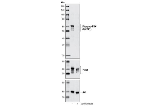
Western blot analysis of extracts from PC3 cells, untreated or λ phosphatase-treated, using Phospho-PDK1 (Ser241) (C49H2) Rabbit mAb (upper), PDK1 Antibody #3062 (middle) or Akt Antibody #9272 (lower).
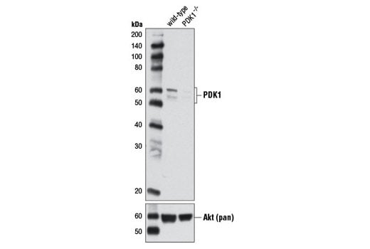
Western blot analysis of extracts from HCT 116 wild-type and HCT 116 PDK1-/- cells using PDK1 (D37A7) Rabbit mAb (upper) and Akt (pan) (C67E7) Rabbit mAb #4691 (lower). (HCT 116 wild-type and HCT 116 PDK1-/- cells were kindly provided by Dr. Bert Vogelstein, Johns Hopkins University, Baltimore, MD).
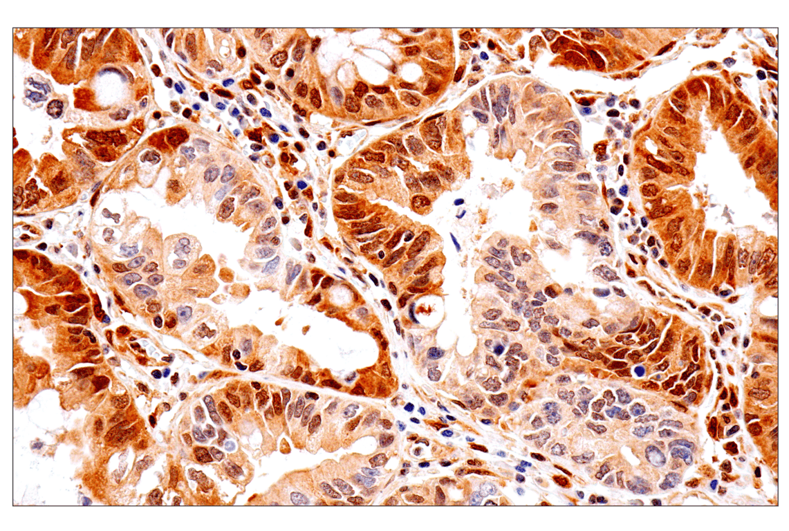
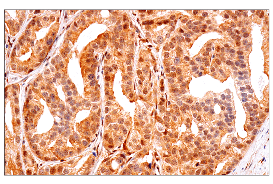
Immunohistochemical analysis of paraffin-embedded human prostate adenocarcinoma using PTEN (D4.3) XP® Rabbit mAb performed on the Leica BOND RX.