


 全部商品分类
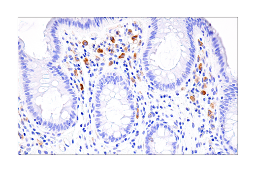


 下载产品说明书 下载SDS
 用小程序,查商品更便捷


 收藏
收藏
 对比
对比 咨询
咨询
The Pyroptosis Antibody Sampler Kit provides an economical means of detecting proteins that are used as readouts for pyroptosis. The kit includes enough antibodies to perform two western blot experiments with each primary antibody.
参考图片
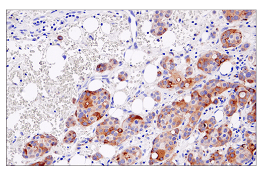
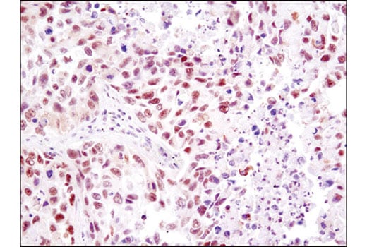
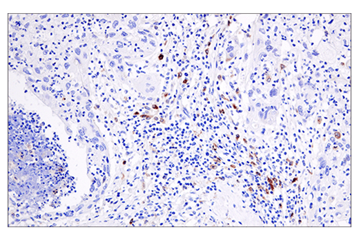
Immunohistochemical analysis of paraffin-embedded human ulcerative colitis using Cleaved Gasdermin D (Asp275) (E7H9G) Rabbit mAb.
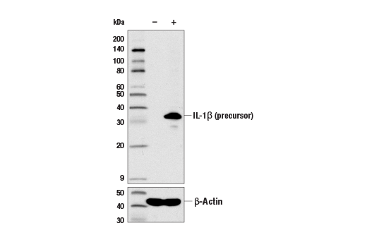
Western blot analysis of extracts from THP-1 cells, untreated (-) or LPS-treated (100 ng/ml, 3 hr; +), using IL-1β (D3U3E) Rabbit mAb (upper) and β-Actin (D6A8) Rabbit mAb #8457 (lower).
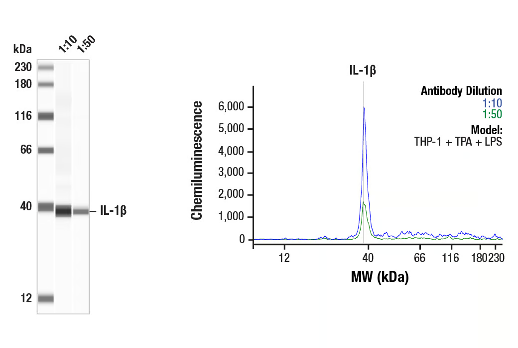
Simple Western™ analysis of lysates (0.1 mg/mL) from THP-1 cells treated with TPA (80nM, O/N) + LPS (1 µg/ml 15min) using IL-1β (D3U3E) Rabbit mAb #12703. The virtual lane view (left) shows the target band (as indicated) at 1:10 and 1:50 dilutions of primary antibody. The corresponding electropherogram view (right) plots chemiluminescence by molecular weight along the capillary at 1:10 (blue line) and 1:50 (green line) dilutions of primary antibody. This experiment was performed under reducing conditions on the Jess™ Simple Western instrument from ProteinSimple, a BioTechne brand, using the 12-230 kDa separation module.
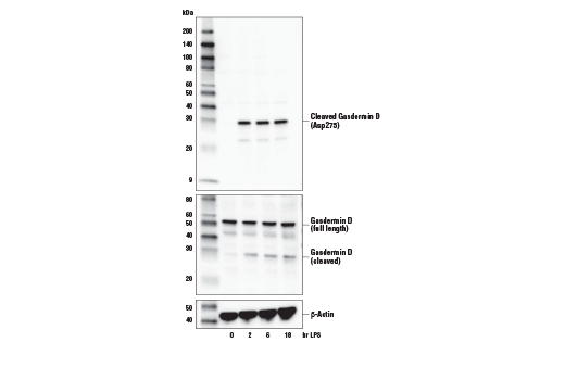
Western blot analysis of extracts from THP-1 cells, differentiated with TPA #4174 (50 ng/ml, overnight) and then treated with LPS #14011 (5 μg/ml, indicated times), using Cleaved Gasdermin D (Asp275) (E7H9G) Rabbit mAb (upper), total Gasdermin D (L60) Antibody #93709 (middle), or β-Actin (D6A8) Rabbit mAb #8457 (lower).
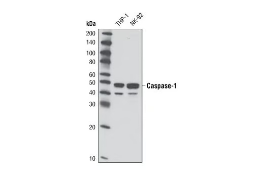
Western blot analysis of extracts from THP-1 and NK-92 cells using Caspase-1 (D7F10) Rabbit mAb.
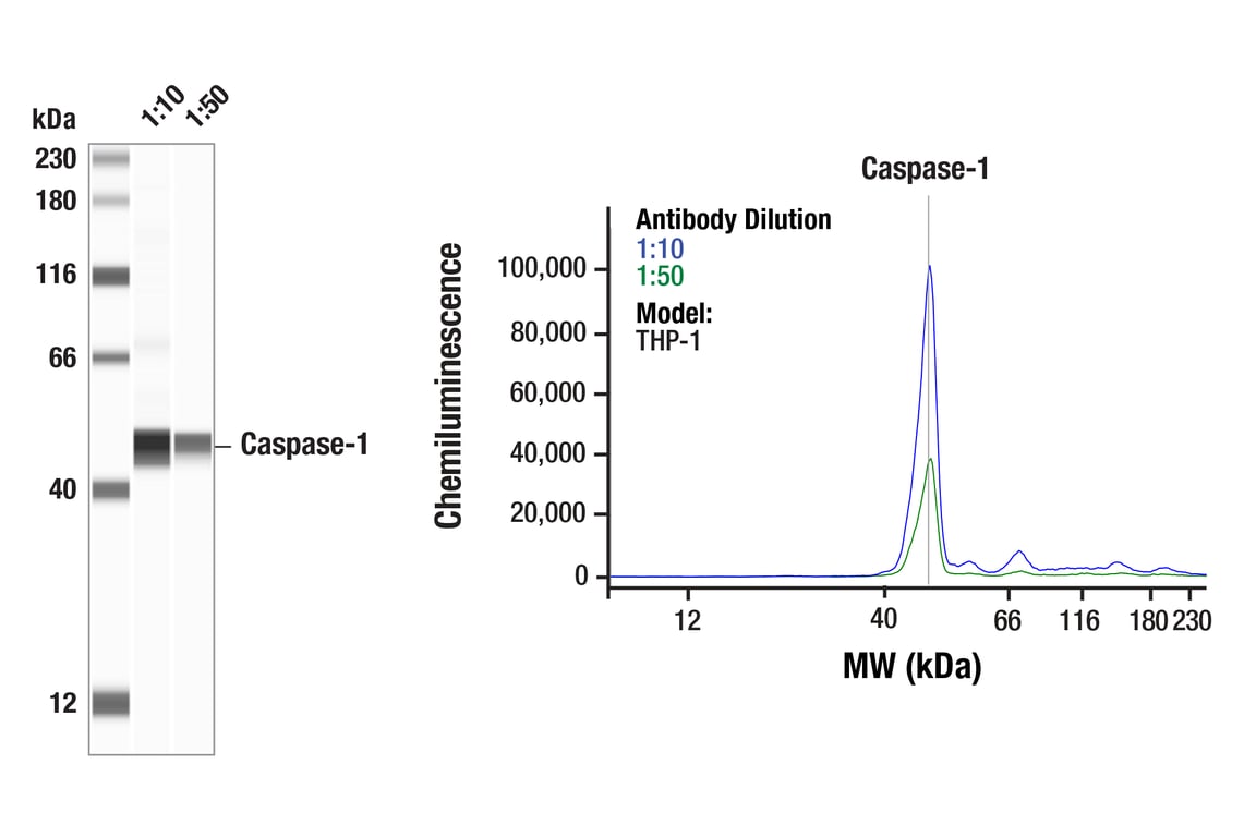
Simple WesternTM analysis of lysates (1.0 mg/mL) from THP-1 cells using Caspase-1 (D7F10) Rabbit mAb #3866. The virtual lane view (left) shows the target band (as indicated) at 1:10 and 1:50 dilutions of primary antibody. The corresponding electropherogram view (right) plots chemiluminescence by molecular weight along the capillary at 1:10 (blue line) and 1:50 (green line) dilutions of primary antibody. This experiment was performed under reducing conditions on the JessTM Simple Western instrument from ProteinSimple, a BioTechne brand, using the 12-230 kDa separation module.
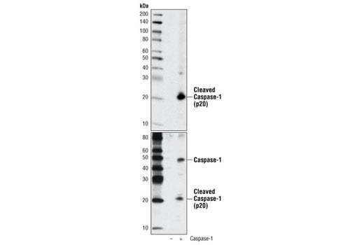
Western blot analysis of extracts from COS-7 cells, untransfected (-) or transfected with construct overexpressing human caspase-1 (+), using Cleaved Caspase-1 (Asp297) (D57A2) Rabbit mAb (upper) or Caspase-1 (D7F10) Rabbit mAb #3866 (lower).
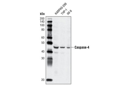
Western blot analysis of extracts from KARPAS-299, THP-1 and IM-9 cells using Caspase-4 Antibody.
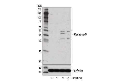
Western blot analysis of extracts from THP-1 cells differentiated with TPA #4174 (80 nM, overnight) followed by treatment with LPS #14011 (1 μg/ml, indicated times), using Caspase-5 (D3G4W) Rabbit mAb (upper) and β-Actin (D6A8) Rabbit mAb #8457 (lower)
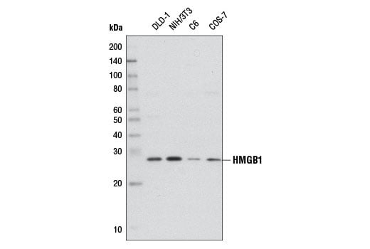
Western blot analysis of extracts from various cell lines using HMGB1 (D3E5) Rabbit mAb.
After the primary antibody is bound to the target protein, a complex with HRP-linked secondary antibody is formed. The LumiGLO® is added and emits light during enzyme catalyzed decomposition.
Western blot analysis of recombinant Human Interleukin-1β (hIL-1β) #8900 using Cleaved-IL-1β (Asp116) (D3A3Z) Rabbit mAb.
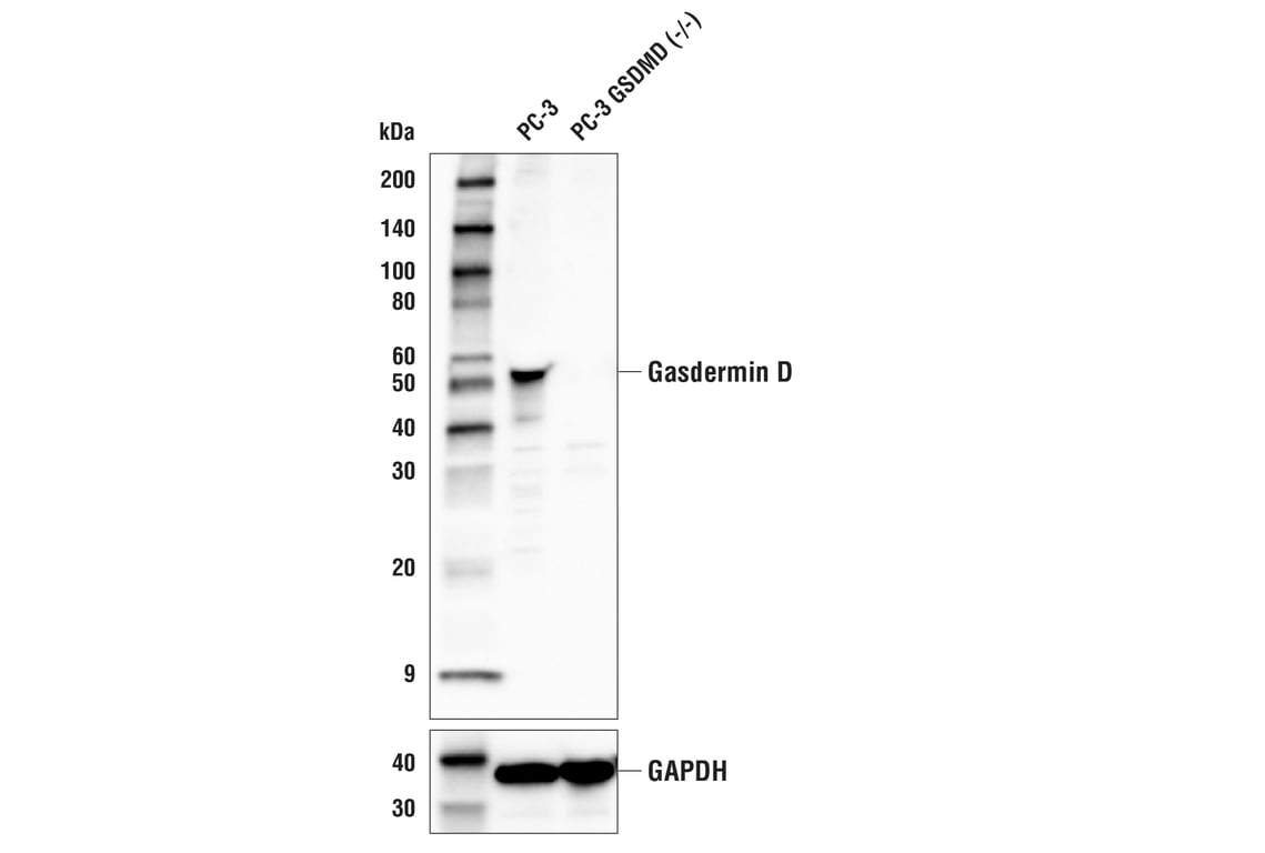
Western blot analysis of extracts from PC-3 cells and PC-3 GSDMD knockout (-/-) cells using Gasdermin D (E8G3F) Rabbit mAb (upper) or GAPDH (D16H11) XP® Rabbit mAb #5174 (lower).
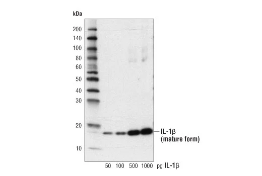
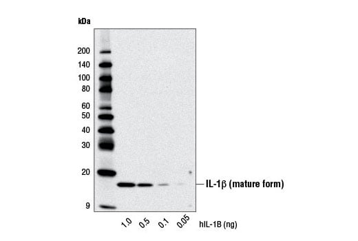
Western blot analysis of recombinant Human Interleukin-1β (hIL-1β) #8900 using IL-1β (D3U3E) Rabbit mAb.
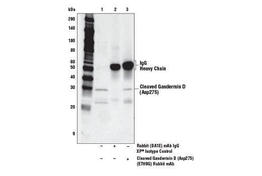
Immunoprecipitation of Cleaved Gasdermin D (Asp725) from THP-1 cells differentiated with TPA #4174 (50 ng/ml, overnight) and then treated with LPS #14011 (5 μg/ml, 6 hr). Lane 1 is 10% input, lane 2 is Rabbit (DA1E) mAb IgG XP® Isotype Control #3900, and lane 3 is Cleaved Gasdermin D (Asp275) (E7H9G) Rabbit mAb. Western blot was performed using Cleaved Gasdermin D (Asp275) (E7H9G) Rabbit mAb. Anti-rabbit IgG, HRP-linked Antibody #7074 was used as a secondary antibody.
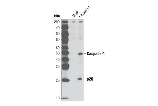
Western blot analysis of extracts from COS cells, untransfected or transfected with human caspase-1, using Caspase-1 (D7F10) Rabbit mAb.
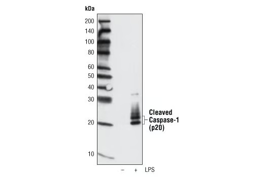
Western blot analysis of extracts from the media of THP-1 cells, differentiated with TPA #9905 (80 nM, overnight) followed by treatment with LPS (1 ug/ml, 8 hours), using Cleaved Caspase-1 (Asp297) (D57A2) Rabbit mAb.
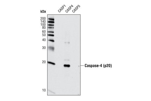
Western blot analyis of extracts from recombinant, active caspase-1, -4, and -5 using Caspase-4 Antibody.
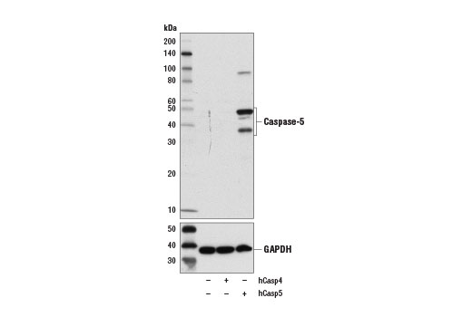
Western blot analysis of extracts form 293T cells, mock transfected (-) or transfected with constructs expressing full-length human caspase-4 protein (hCasp4; +) or caspase-5 protein (hCasp5; +), using Caspase-5 (D3G4W) Rabbit mAb (upper) and GAPDH (D16H11) XP® Rabbit mAb #5174 (lower).
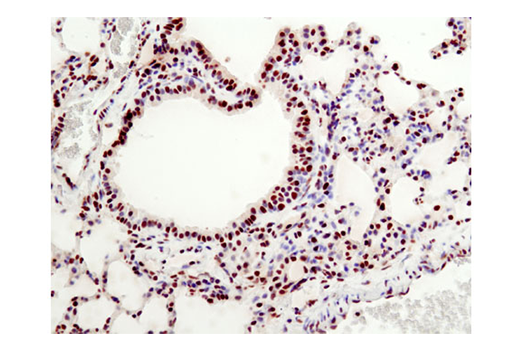
Immunohistochemical analysis of paraffin-embedded mouse lung using HMGB1 (D3E5) Rabbit mAb.
Western blot analysis of extracts from the media of mouse bone marrow derived macrophages (mBMDM), untreated (-) or treated with Lipopolysaccharides (LPS) #14011 (50 ng/ml, 4 hr; +) followed by Nigericin (15 μM, 45 min; +), using HMGB1 (D3E5) Rabbit mAb.
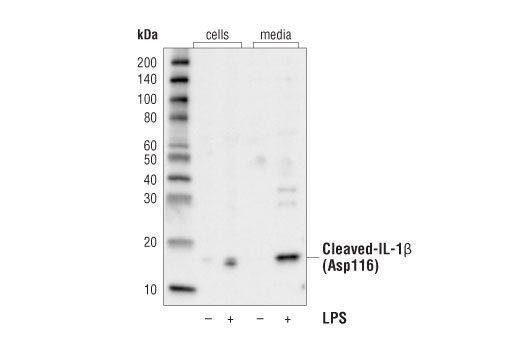
Western blot analysis of extracts from cells or media collected from THP-1 cells, differentiated with TPA #4147 (80 nM, overnight) and subsequently treated with (+) or without (-) Lipopolysaccharides (LPS) #14011 (1 μg/ml, 6 hr), using Cleaved-IL-1β (Asp116) (D3A3Z) Rabbit mAb.
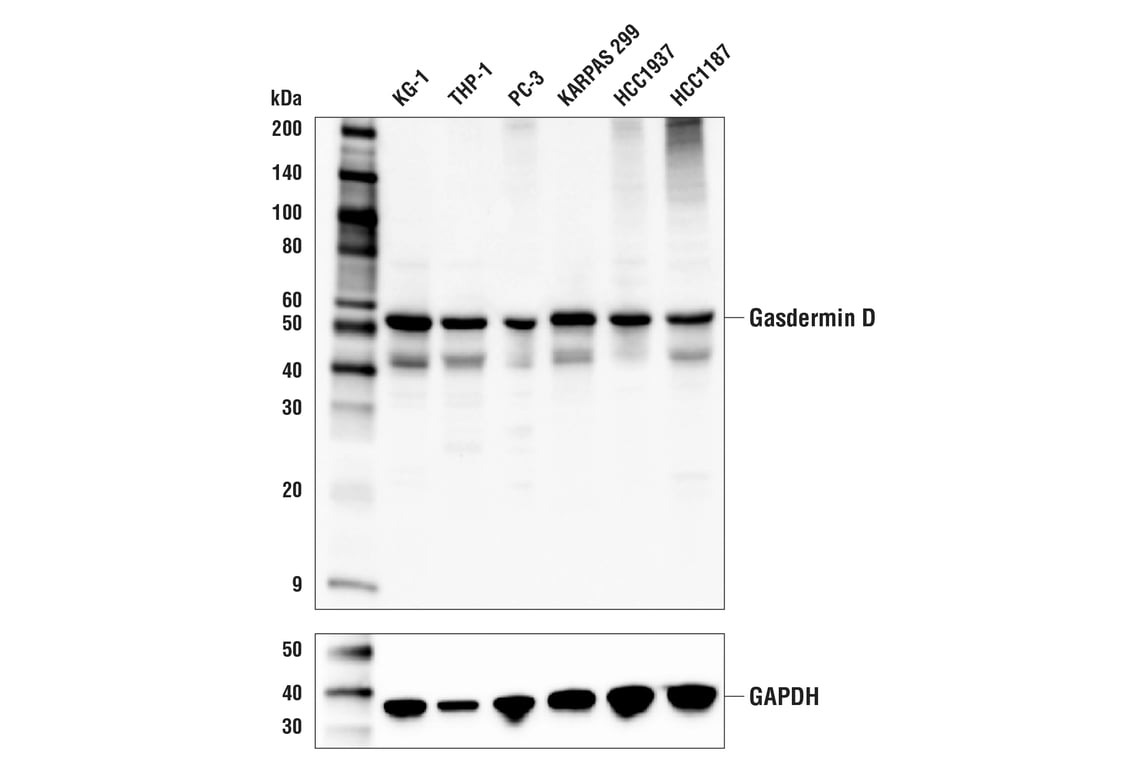
Western blot analysis of extracts from various cell lines using Gasdermin D (E8G3F) Rabbit mAb (upper) or GAPDH (D16H11) XP® Rabbit mAb #5174 (lower).
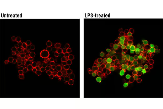
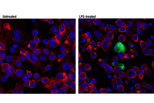
Confocal immunofluorescent analysis of THP-1 cells, untreated (left) or LPS-treated (500 ng/ml, 2 hr; right), using IL-1β (D3U3E) Rabbit mAb (green). Actin filaments were labeled with DY-554 phalloidin (red).
Immunohistochemical analysis of paraffin-embedded human ductal breast carcinoma using Cleaved Gasdermin D (Asp275) (E7H9G) Rabbit mAb.
Immunohistochemical analysis of paraffin-embedded human colon carcioma using HMGB1 (D3E5) Rabbit mAb.
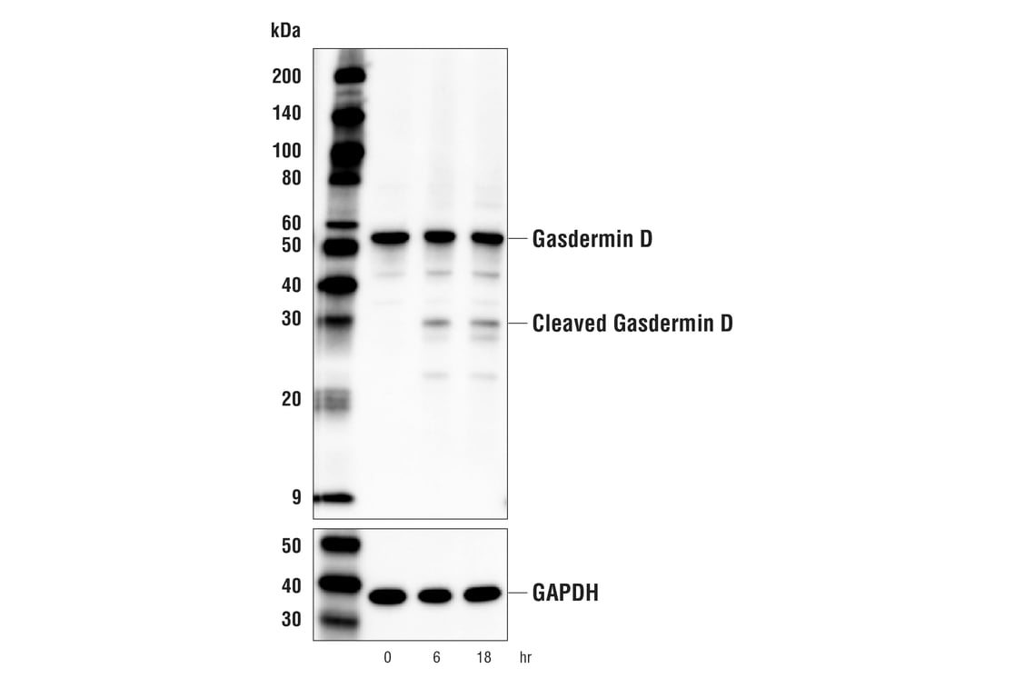
Western blot analysis of extracts from THP-1 cells, differentiated with TPA #4174 (50 ng/ml, overnight) and then treated with LPS #14011 (5 μg/ml, indicated times), using Gasdermin D (E8G3F) Rabbit mAb (upper) or GAPDH (D16H11) XP® Rabbit mAb #5174 (lower).
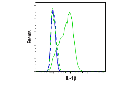
Flow cytometric analysis of THP-1, untreated (blue, negative) or treated with LPS #14011 (100 ng/ml, 3 hr; green, positive) using IL-1β (D3U3E) Rabbit mAb (solid lines) or concentration-matched Rabbit (DA1E) mAb IgG XP® Isotype Control #3900 (dashed lines). Anti-rabbit IgG (H+L), F(ab')₂ Fragment (Alexa Fluor® 488 Conjugate) #4412 was used as a secondary antibody.
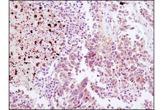
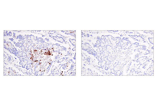
Immunohistochemical analysis of paraffin-embedded human colon carcinoma using Cleaved Gasdermin D (Asp275) (E7H9G) Rabbit mAb in the presence of non-cleaved Gasdermin D peptide (left) or Asp275 cleavage-specific Gasdermin D peptide (right).
Immunohistochemical analysis of paraffin-embedded human lung carcioma using HMGB1 (D3E5) Rabbit mAb.
Confocal immunofluorescent analysis of THP-1 cells, differentiated with TPA #4174 (80 nM, 24 hr) and subsequently treated with (right) or without (left) Lipopolysaccharides (LPS) #14011 (1 μg/ml, 6 hr), using Cleaved-IL-1β (Asp116) (D3A3Z) Rabbit mAb (green). Actin filaments were labeled with DyLight™ 554 Phalloidin #13054 (red). Blue pseudocolor = DRAQ5® #4084 (fluorescent DNA dye).
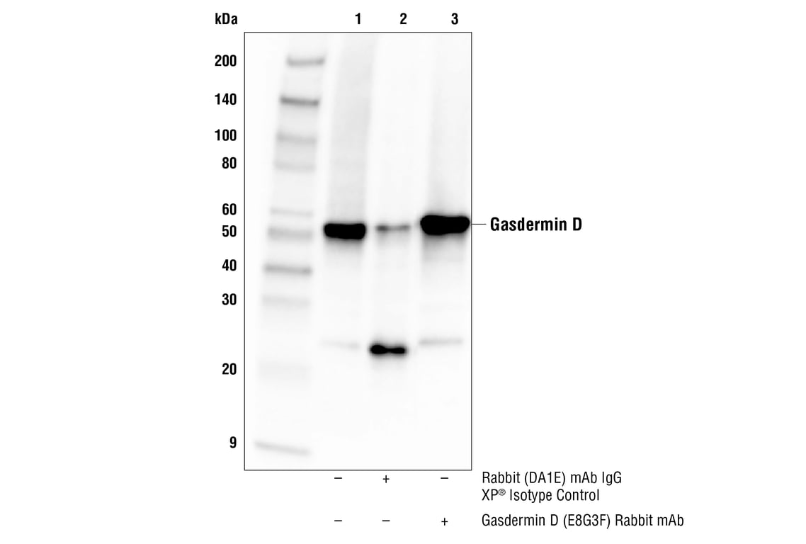
Immunoprecipitation of Gasdermin D protein from THP-1 cell extracts. Lane 1 is 10% input, lane 2 is Rabbit (DA1E) mAb IgG XP® Isotype Control #3900, and lane 3 is Gasdermin D (E8G3F) Rabbit mAb. Western blot analysis was performed using Gasdermin D (E8G3F) Rabbit mAb. Mouse Anti-rabbit IgG (Conformation Specific) (L27A9) mAb (HRP Conjugate) #5127 was used as a secondary antibody.
Immunohistochemical analysis of paraffin-embedded human squamous cell lung carcinoma using Cleaved Gasdermin D (Asp275) (E7H9G) Rabbit mAb.
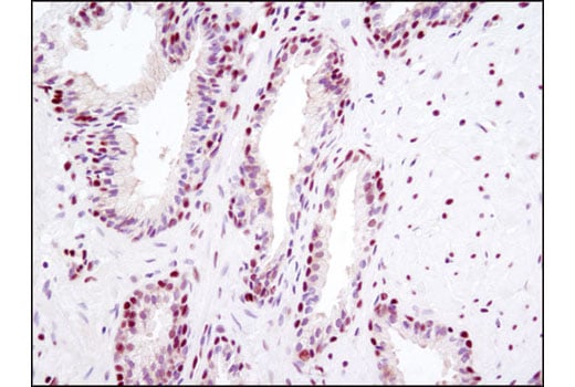
Immunohistochemical analysis of paraffin-embedded human prostate carcioma using HMGB1 (D3E5) Rabbit mAb.
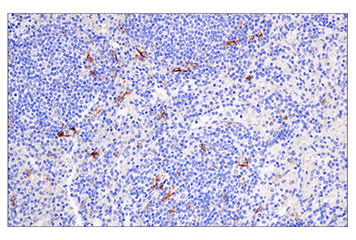
Immunohistochemical analysis of paraffin-embedded human non-Hodgkin's Lymphoma using Cleaved Gasdermin D (Asp275) (E7H9G) Rabbit mAb.
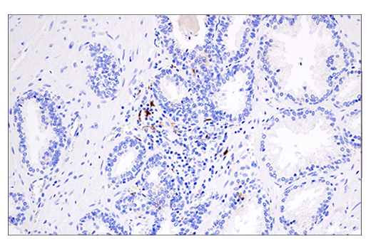
Immunohistochemical analysis of paraffin-embedded human prostate carcinoma using Cleaved Gasdermin D (Asp275) (E7H9G) Rabbit mAb.
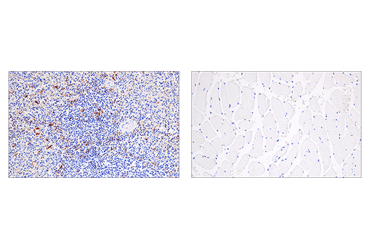
Immunohistochemical analysis of paraffin-embedded human spleen (left, positive) and skeletal muscle (right, negative) using Cleaved Gasdermin D (Asp275) (E7H9G) Rabbit mAb.
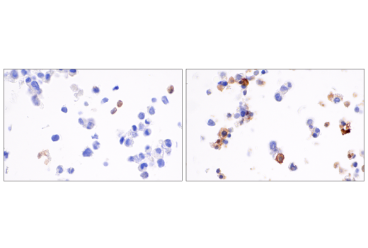
Immunohistochemical analysis of paraffin-embedded THP-1 cell pellets, differentiated with TPA #4174 (left) and then treated with LPS #14011 (right), using Cleaved Gasdermin D (Asp275) (E7H9G) Rabbit mAb.