

 全部商品分类
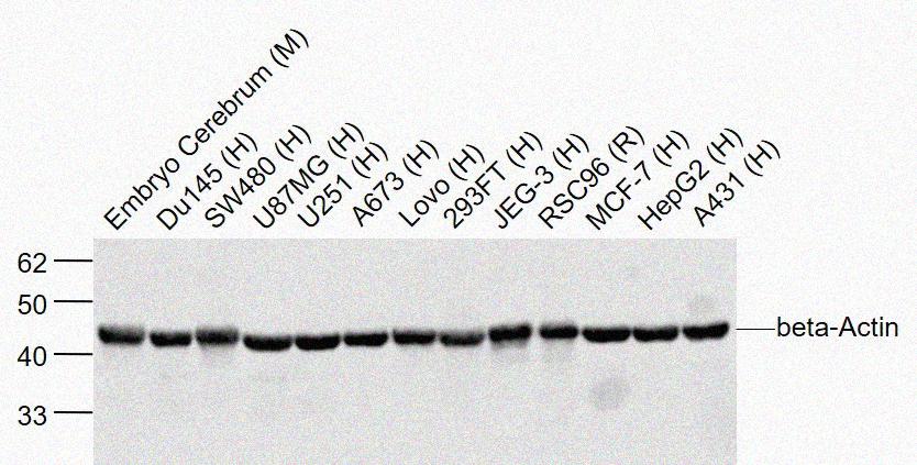


 下载产品说明书
 用小程序,查商品更便捷


 收藏
收藏
 对比
对比 咨询
咨询Disclaimer note: The observed molecular weight of the protein may vary from the listed predicted molecular weight due to post translational modifications, post translation cleavages, relative charges, and other experimental factors.

Disclaimer note: The observed molecular weight of the protein may vary from the listed predicted molecular weight due to post translational modifications, post translation cleavages, relative charges, and other experimental factors.
参考图片
Sample:Embryo Cerebrum (Mouse) Lysate at 40 ug Du145 (Human) Lysate at 40 ug SW480 (Human) Cell Lysate at 40 ug U87MG (Human) Lysate at 40 ug U251 (Human) Lysate at 40 ug A673 (Human) Lysate at 40 ug Lovo (Human) Lysate at 40 ug 293FT (Human) Lysate at 40 ug JEG-3 (Human) Lysate at 40 ug RSC96 (Rat) Cell Lysate at 40 ug MCF-7 (Human) Cell Lysate at 40 ug HepG2 (Human) Lysate at 40 ug A431 (Human) Lysate at 40 ug Primary: Anti-beta-Actin at 1/2000 dilution Secondary: IRDye800CW Goat Anti-Rabbit IgG at 1/20000 dilution Predicted band size: 42 kD Observed band size: 42 kD
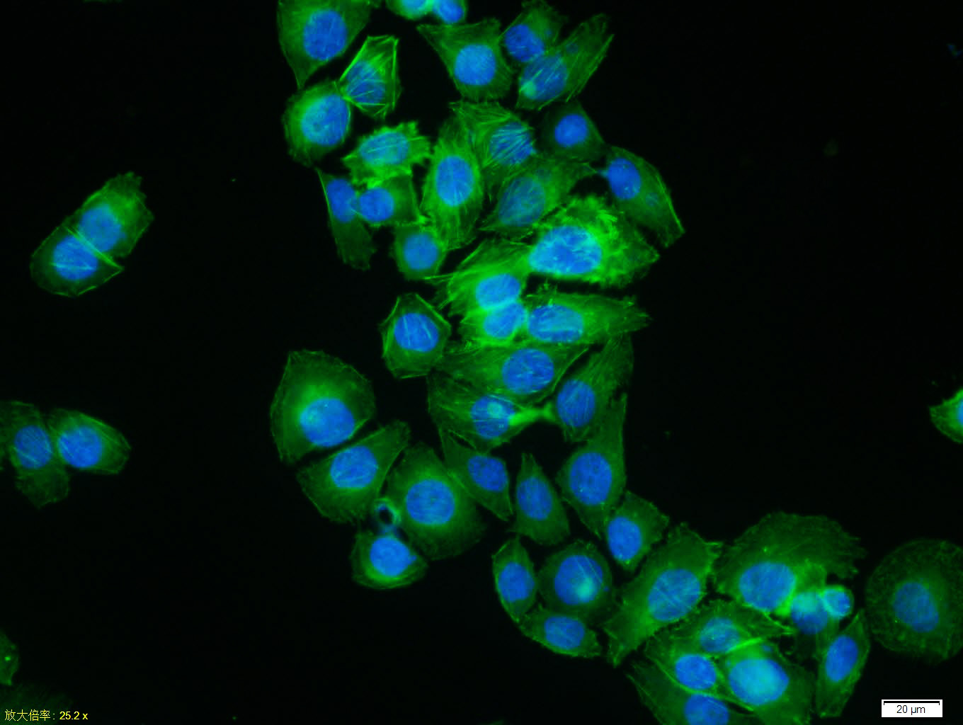
MCF7 cell; 4% Paraformaldehyde-fixed; Triton X-100 at room temperature for 20 min; Blocking buffer (normal goat serum) at 37°C for 20 min; Antibody incubation with (beta-Actin) polyclonal Antibody, Unconjugated 1:100, 90 minutes at 37°C; followed by a conjugated Goat Anti-Rabbit IgG antibody at 37°C for 90 minutes, DAPI (blue) was used to stain the cell nuclei.
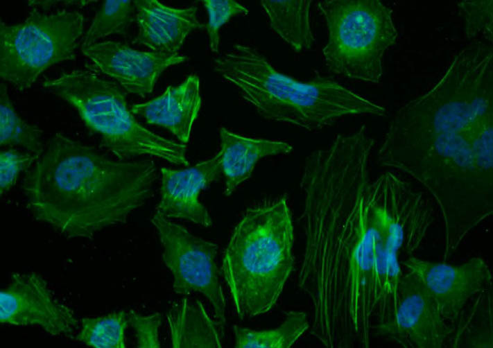
Tissue/cell: Hela cell; 4% Paraformaldehyde-fixed; Triton X-100 at room temperature for 20 min; Blocking buffer (normal goat serum) at 37°C for 20 min; Antibody incubation with (beta-Actin) polyclonal Antibody, Unconjugated 1:100, 90 minutes at 37°C; followed by a conjugated Goat Anti-Rabbit IgG-FITC antibody at 37°C for 90 minutes, DAPI (blue) was used to stain the cell nuclei.
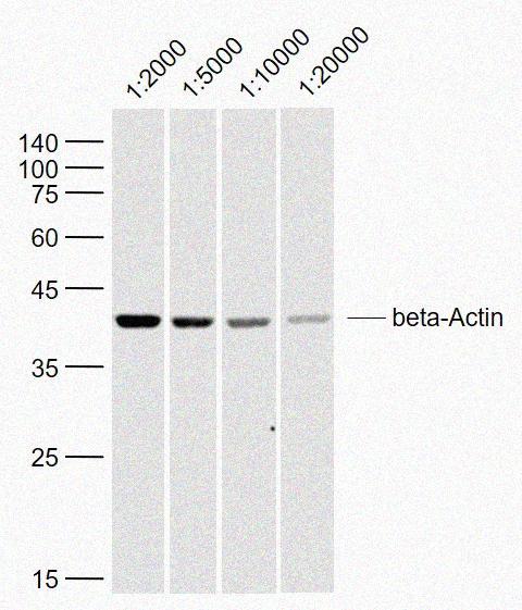
Sample: SH-SY5Y (Human) Lysate at 40 ug Primary: Anti-beta-Actin at 1/2000~1/20000 dilution Secondary: IRDye800CW Goat Anti-Rabbit IgG at 1/20000 dilution Predicted band size: 42 kD Observed band size: 42 kD
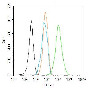
Blank control: NIH/3T3. Primary Antibody (green line): Rabbit Anti-beta-Actin (Loading Control) antibody Dilution: 1ug/10^6 cells; Isotype Control Antibody (orange line): Rabbit IgG. Secondary Antibody : Goat anti-rabbit IgG-AF488 Dilution: 1ug/T. Protocol The cells were fixed with 4% PFA (10min at room temperature)and then permeabilized with 90% ice-cold methanol for 20 min at -20℃. The cells were then incubated in 5% BSA to block non-specific protein-protein interactions for 30 min at room temperature.Cells stained with Primary Antibody for 30 min at room temperature. The secondary antibody used for 40 min at room temperature. Acquisition of 20,000 events was performed.