


 全部商品分类
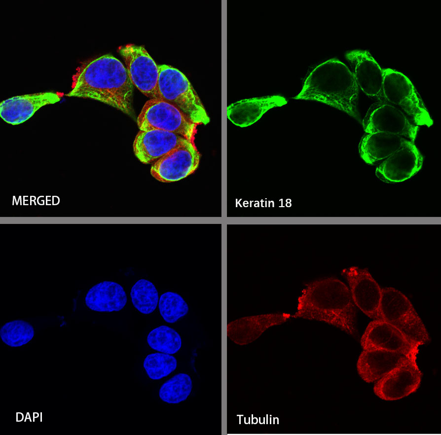


 下载产品说明书
 用小程序,查商品更便捷


 收藏
收藏
 对比
对比 咨询
咨询Disclaimer note: The observed molecular weight of the protein may vary from the listed predicted molecular weight due to post translational modifications, post translation cleavages, relative charges, and other experimental factors.

K18 a type I cytoskeletal keratin. The keratins are intermediate filament proteins responsible for the structural integrity of epithelial cells and are subdivided into cytokeratins and hair keratins. Keratin 18 and its filament partner keratin 8 are perhaps the most commonly found members of the intermediate filament gene family. They are expressed in single layer epithelial tissues of the body.
Disclaimer note: The observed molecular weight of the protein may vary from the listed predicted molecular weight due to post translational modifications, post translation cleavages, relative charges, and other experimental factors.
参考图片
IHC shows positive staining in paraffin-embedded rat liver. Anti-Keratin 18 antibody was used at 1/2000 dilution, followed by a HRP Polymer for Mouse & Rabbit IgG (ready to use). Counterstained with hematoxylin. Heat mediated antigen retrieval with Tris/EDTA buffer pH9.0 was performed before commencing with IHC staining protocol.
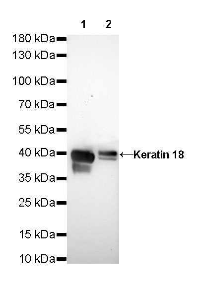
WB resuLt of Keratin 18 Rabbit mAb
Primary antibody: Keratin 18 Rabbit mAb at 1/1000 dilution
Lane 1: MCF7 whole cell lysate 20 µg
Lane 2: HeLa whole cell lysate 20 µg
Secondary antibody: Goat Anti-Rabbit IgG, (H+L), HRP conjugated at 1/10000 dilution
Predicted MW: 46 kDa
Observed MW: 42 kDa
Exposure time: 6s
Negative tissue: IHC shows negative staining in paraffin-embedded human cervical squamous cell carcinoma. Anti-Keratin 18 antibody was used at 1/2000 dilution, followed by a HRP Polymer for Mouse & Rabbit IgG (ready to use). Counterstained with hematoxylin. Heat mediated antigen retrieval with Tris/EDTA buffer pH9.0 was performed before commencing with IHC staining protocol.
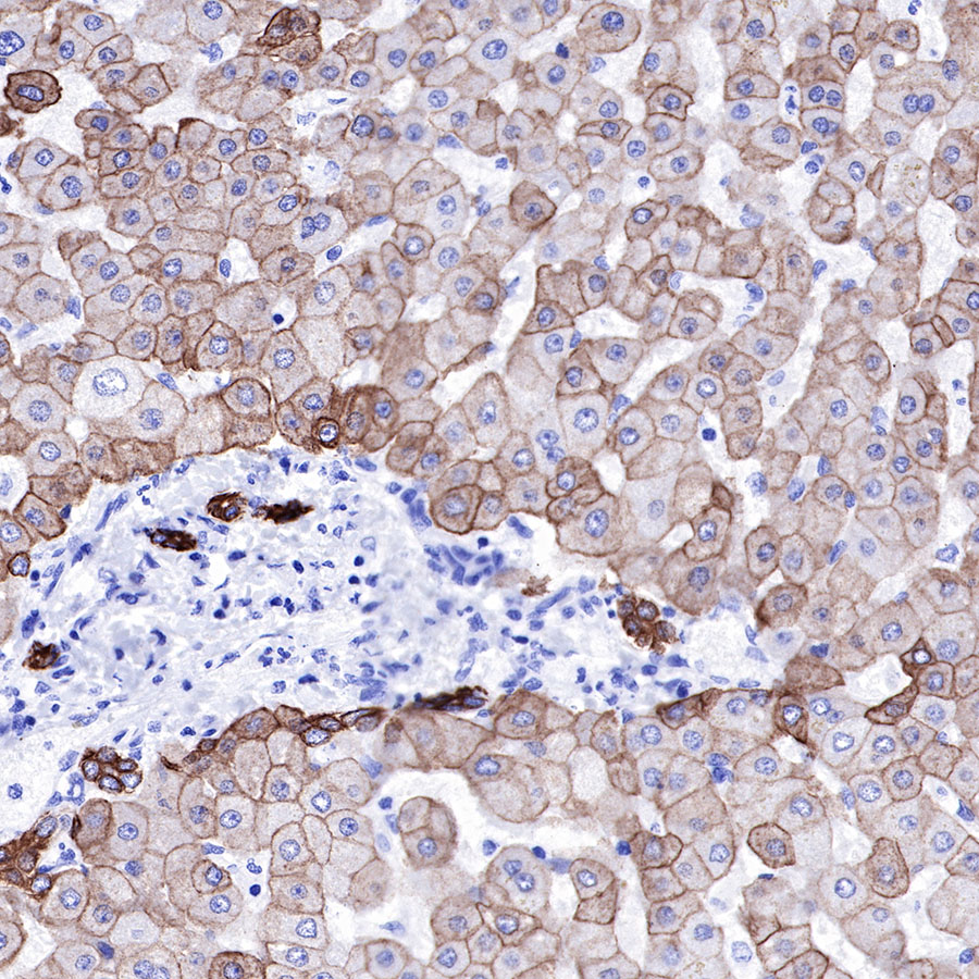
IHC shows positive staining in paraffin-embedded mouse liver. Anti-Keratin 18 antibody was used at 1/2000 dilution, followed by a HRP Polymer for Mouse & Rabbit IgG (ready to use). Counterstained with hematoxylin. Heat mediated antigen retrieval with Tris/EDTA buffer pH9.0 was performed before commencing with IHC staining protocol.
IHC shows positive staining in paraffin-embedded human ovarian carcinoma. Anti-Keratin 18 antibody was used at 1/2000 dilution, followed by a HRP Polymer for Mouse & Rabbit IgG (ready to use). Counterstained with hematoxylin. Heat mediated antigen retrieval with Tris/EDTA buffer pH9.0 was performed before commencing with IHC staining protocol.
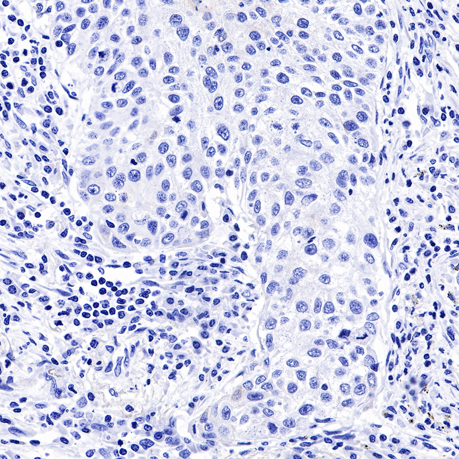
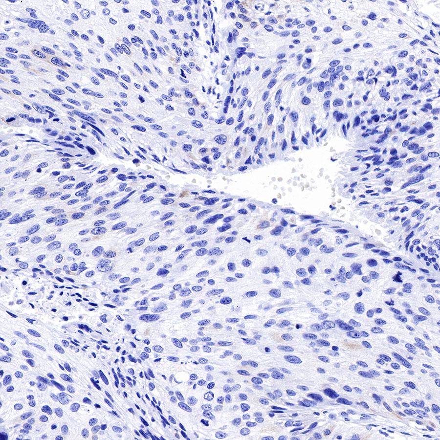
Negative tissue: IHC shows negative staining in paraffin-embedded human lung squamous cell carcinoma. Anti-Keratin 18 antibody was used at 1/2000 dilution, followed by a HRP Polymer for Mouse & Rabbit IgG (ready to use). Counterstained with hematoxylin. Heat mediated antigen retrieval with Tris/EDTA buffer pH9.0 was performed before commencing with IHC staining protocol.
ICC shows positive staining in MCF7 cells. Anti-Keratin 18 antibody was used at 1/50 dilution (Green) and incubated overnight at 4°C. Goat polyclonal Antibody to Rabbit IgG - H&L (Alexa Fluor® 488) was used as secondary antibody at 1/1000 dilution. The cells were fixed with 4% PFA and permeabilized with 0.1% PBS-Triton X-100. Nuclei were counterstained with DAPI (Blue). Counterstain with tubuLin (red).
Flow cytometric analysis of 4% PFA fixed 90% methanol permeabilized MCF7 (Human breast adenocarcinoma epithelial cell) labelling Keratin 18 antibody at 1/50 dilution (0.1 ug) / (Red) compared with a Rabbit monoclonal IgG (Black) isotype control and an unlabelled control (cells without incubation with primary antibody and secondary antibody) (Blue). Goat Anti - Rabbit IgG Alexa Fluor® 488 was used as the secondary antibody.
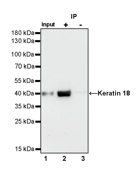
Keratin 18 Rabbit mAb at 1/25 dilution (0.2 µg) immunoprecipitating Keratin 18 in 0.2 mg HeLa whole cell lysate.
Western blot was performed on the immunoprecipitate using Keratin 18 Rabbit mAb at 1/1000 dilution.
Secondary antibody (HRP) for IP was used at 1/400 dilution.
Lane 1: HeLa whole cell lysate 5 µg (Input)
Lane 2: Keratin 18 Rabbit mAb IP in HeLa whole cell lysate
Lane 3: Rabbit monoclonal IgG IP in HeLa whole cell lysate
Predicted MW: 46 kDa
Observed MW: 42 kDa
Exposure time: 30 s
WB result of Keratin 18 Rabbit mAb Primary antibody: Keratin 18 Rabbit mAb at 1/1000 dilution Lane 1: MCF7 whole cell lysate 20 µg Lane 2: HeLa whole cell lysate 20 µg Secondary antibody: #abs20040 at 1/10000 dilution Predicted MW: 46 kDa Observed MW: 42 kDa Exposure time: 6s
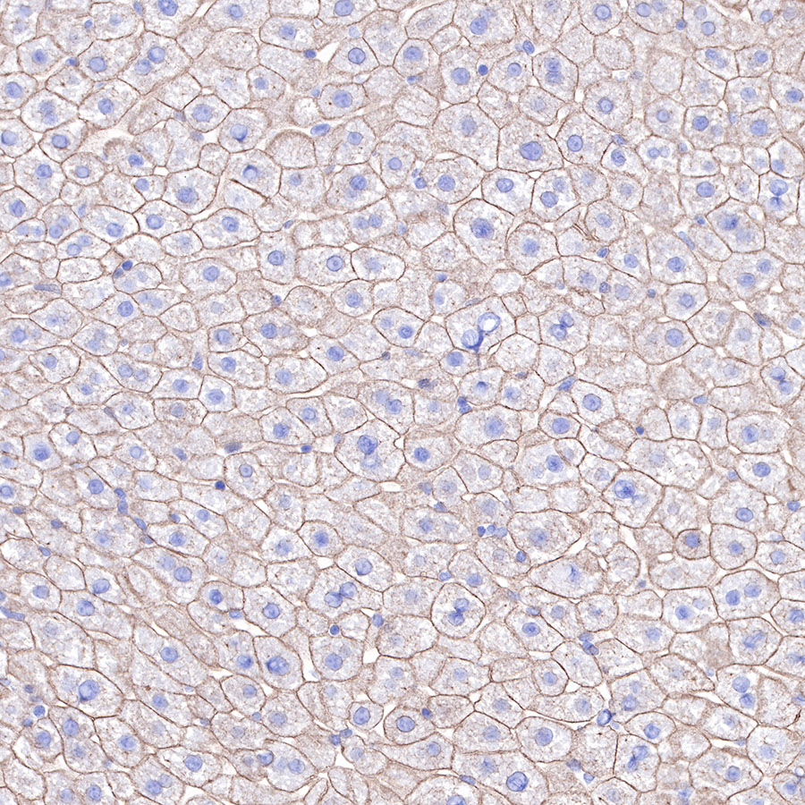
IHC shows positive staining in paraffin-embedded human liver. Anti-Keratin 18 antibody was used at 1/2000 dilution, Secondary antibody: #abs20040. Counterstained with hematoxylin. Heat mediated antigen retrieval with Tris/EDTA buffer pH9.0 was performed before commencing with IHC staining protocol.
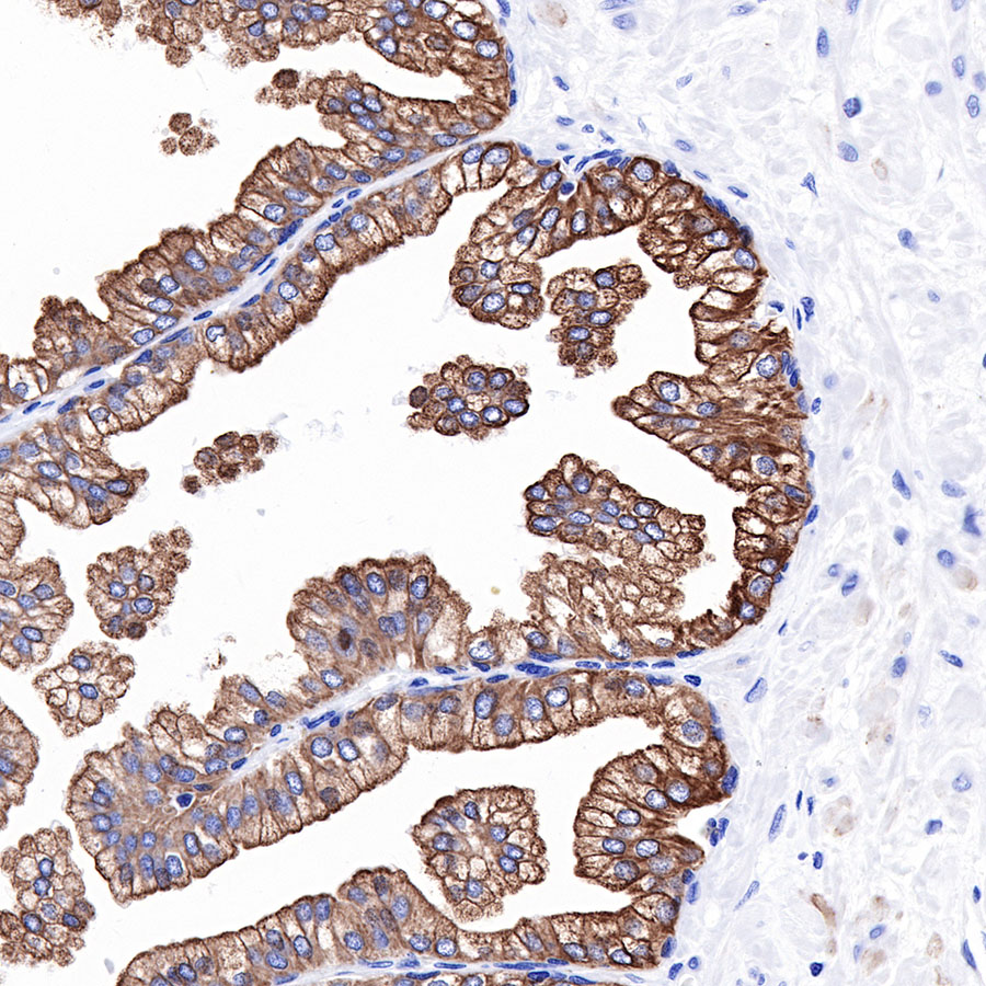
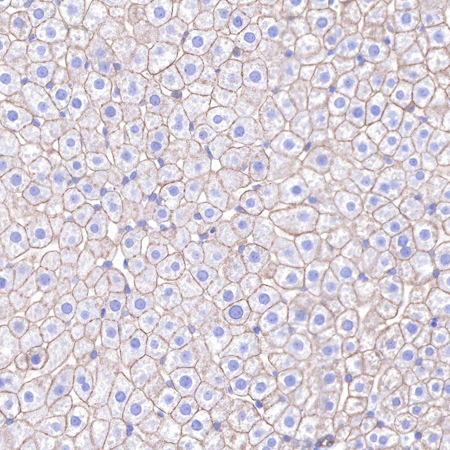
IHC shows positive staining in paraffin-embedded human prostatic hyperplasia. Anti-Keratin 18 antibody was used at 1/2000 dilution, Secondary antibody: #abs20040. Counterstained with hematoxylin. Heat mediated antigen retrieval with Tris/EDTA buffer pH9.0 was performed before commencing with IHC staining protocol.
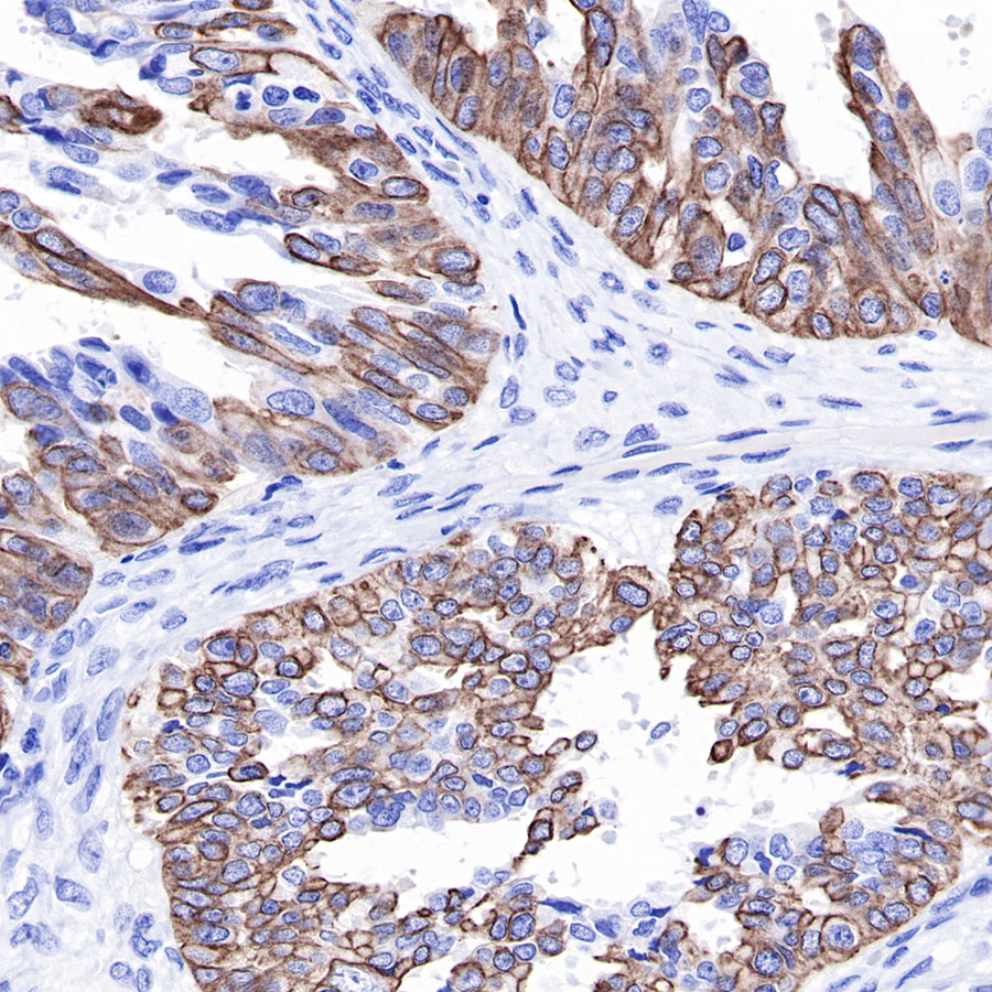
IHC shows positive staining in paraffin-embedded human lung adenocarcinoma. Anti-Keratin 18 antibody was used at 1/2000 dilution, Secondary antibody: #abs20040. Counterstained with hematoxylin. Heat mediated antigen retrieval with Tris/EDTA buffer pH9.0 was performed before commencing with IHC staining protocol.
IHC shows positive staining in paraffin-embedded human ovarian carcinoma. Anti-Keratin 18 antibody was used at 1/2000 dilution, Secondary antibody: #abs20040. Counterstained with hematoxylin. Heat mediated antigen retrieval with Tris/EDTA buffer pH9.0 was performed before commencing with IHC staining protocol.
Negative tissue: IHC shows negative staining in paraffin-embedded human lung squamous cell carcinoma. Anti-Keratin 18 antibody was used at 1/2000 dilution, Secondary antibody: #abs20040. Counterstained with hematoxylin. Heat mediated antigen retrieval with Tris/EDTA buffer pH9.0 was performed before commencing with IHC staining protocol.
Negative tissue: IHC shows negative staining in paraffin-embedded human cervical squamous cell carcinoma. Anti-Keratin 18 antibody was used at 1/2000 dilution, Secondary antibody: #abs20040. Counterstained with hematoxylin. Heat mediated antigen retrieval with Tris/EDTA buffer pH9.0 was performed before commencing with IHC staining protocol.
IHC shows positive staining in paraffin-embedded mouse liver. Anti-Keratin 18 antibody was used at 1/2000 dilution, Secondary antibody: #abs20040. Counterstained with hematoxylin. Heat mediated antigen retrieval with Tris/EDTA buffer pH9.0 was performed before commencing with IHC staining protocol.
IHC shows positive staining in paraffin-embedded rat liver. Anti-Keratin 18 antibody was used at 1/2000 dilution, Secondary antibody: #abs20040. Counterstained with hematoxylin. Heat mediated antigen retrieval with Tris/EDTA buffer pH9.0 was performed before commencing with IHC staining protocol.
IHC shows positive staining in paraffin-embedded human liver. Anti-Keratin 18 antibody was used at 1/2000 dilution, followed by a HRP Polymer for Mouse & Rabbit IgG (ready to use). Counterstained with hematoxylin. Heat mediated antigen retrieval with Tris/EDTA buffer pH9.0 was performed before commencing with IHC staining protocol.
IHC shows positive staining in paraffin-embedded human prostatic hyperplasia. Anti-Keratin 18 antibody was used at 1/2000 dilution, followed by a HRP Polymer for Mouse & Rabbit IgG (ready to use). Counterstained with hematoxylin. Heat mediated antigen retrieval with Tris/EDTA buffer pH9.0 was performed before commencing with IHC staining protocol.
IHC shows positive staining in paraffin-embedded human lung adenocarcinoma. Anti-Keratin 18 antibody was used at 1/2000 dilution, followed by a HRP Polymer for Mouse & Rabbit IgG (ready to use). Counterstained with hematoxylin. Heat mediated antigen retrieval with Tris/EDTA buffer pH9.0 was performed before commencing with IHC staining protocol.