


 全部商品分类
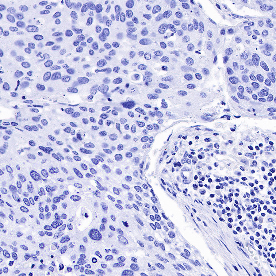


 用小程序,查商品更便捷


参考图片
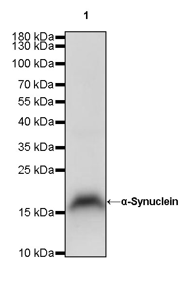
WB result of α-Synuclein Rabbit mAb Primary antibody: α-Synuclein Rabbit mAb at 1/1000 dilution Lane 1: mouse brain lysate 20 ug Secondary antibody: Goat Anti-Rabbit IgG, (H+L), HRP conjugated at 1/10000 dilution Predicted MW: 14 kDa Observed MW: 18 kDa Exposure time: 120 s
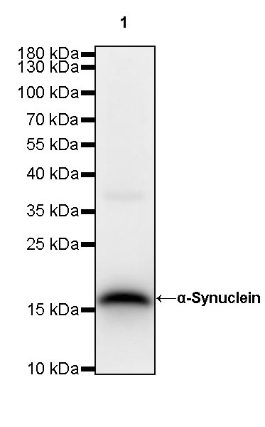
WB result of α-Synuclein Rabbit mAb Primary antibody: α-Synuclein Rabbit mAb at 1/1000 dilution Lane 1: rat brain lysate 20 ug Secondary antibody: Goat Anti-Rabbit IgG, (H+L), HRP conjugated at 1/10000 dilution Predicted MW: 14 kDa Observed MW: 18 kDa Exposure time: 180 s
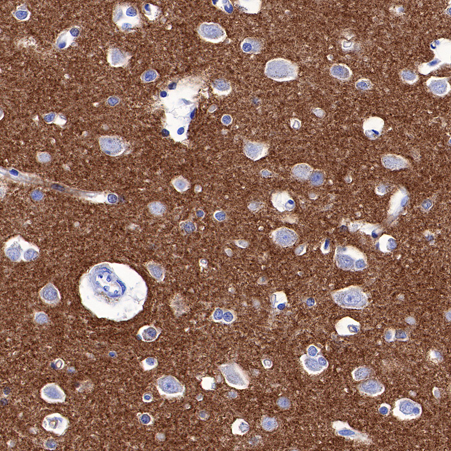
IHC shows positive staining in paraffin-embedded human cerebral cortex. Anti-α-Synuclein antibody was used at 1/2000 dilution, followed by a HRP Polymer for Mouse & Rabbit IgG (ready to use). Counterstained with hematoxylin. Heat mediated antigen retrieval with Tris/EDTA buffer pH9.0 was performed before commencing with IHC staining protocol.
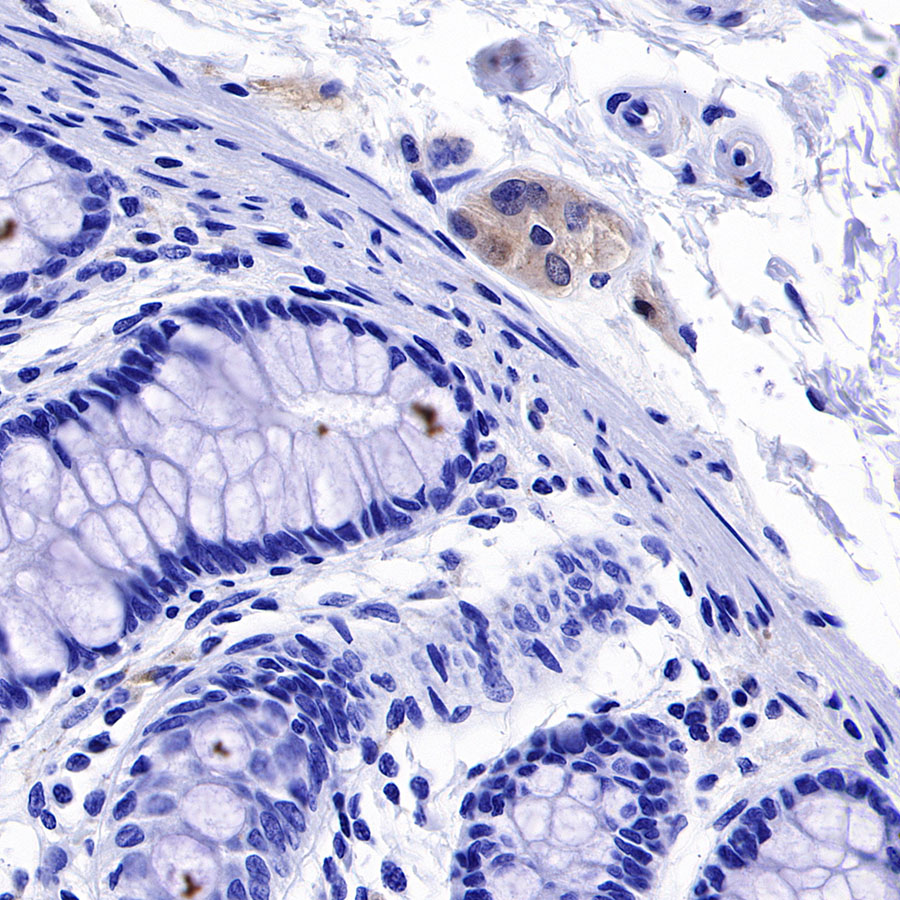
IHC shows positive staining in paraffin-embedded human colon. Anti-α-Synuclein antibody was used at 1/2000 dilution, followed by a HRP Polymer for Mouse & Rabbit IgG (ready to use). Counterstained with hematoxylin. Heat mediated antigen retrieval with Tris/EDTA buffer pH9.0 was performed before commencing with IHC staining protocol.
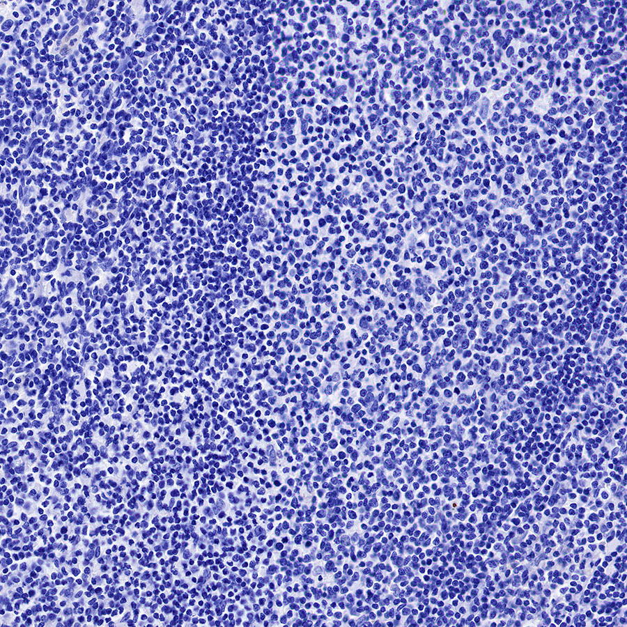
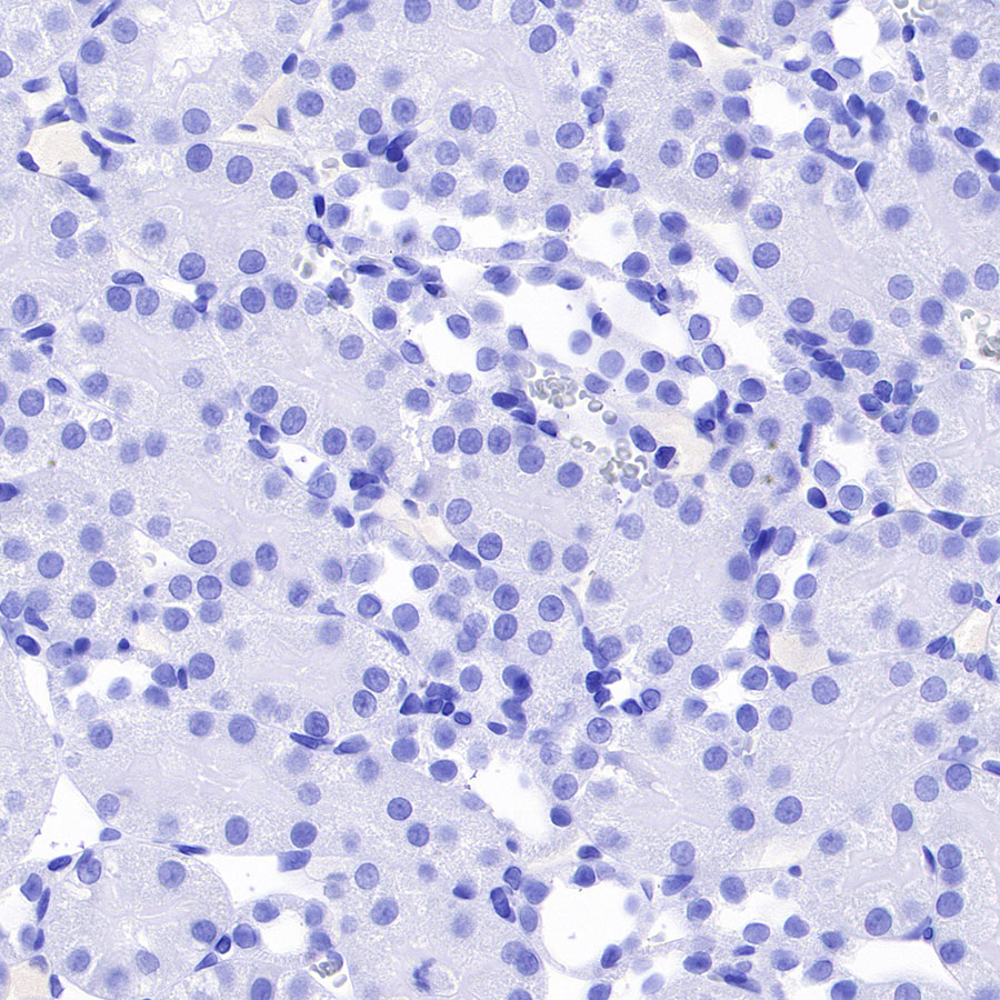
Negative control: IHC shows negative staining in paraffin-embedded human tonsil. Anti-α-Synuclein antibody was used at 1/2000 dilution, followed by a HRP Polymer for Mouse & Rabbit IgG (ready to use). Counterstained with hematoxylin. Heat mediated antigen retrieval with Tris/EDTA buffer pH9.0 was performed before commencing with IHC staining protocol.
Negative control: IHC shows negative staining in paraffin-embedded human cervical squamous cell carcinoma. Anti-α-Synuclein antibody was used at 1/2000 dilution, followed by a HRP Polymer for Mouse & Rabbit IgG (ready to use). Counterstained with hematoxylin. Heat mediated antigen retrieval with Tris/EDTA buffer pH9.0 was performed before commencing with IHC staining protocol.
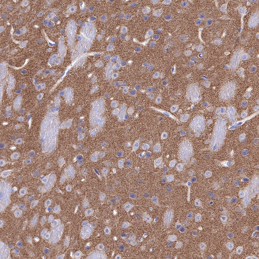
IHC shows positive staining in paraffin-embedded mouse cerebral cortex. Anti-α-Synuclein antibody was used at 1/2000 dilution, followed by a HRP Polymer for Mouse & Rabbit IgG (ready to use). Counterstained with hematoxylin. Heat mediated antigen retrieval with Tris/EDTA buffer pH9.0 was performed before commencing with IHC staining protocol.
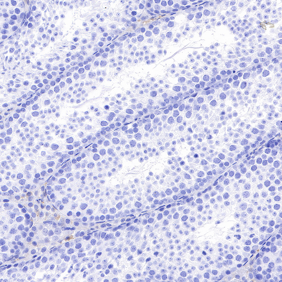
Negative control: IHC shows negative staining in paraffin-embedded mouse testis. Anti-α-Synuclein antibody was used at 1/2000 dilution, followed by a HRP Polymer for Mouse & Rabbit IgG (ready to use). Counterstained with hematoxylin. Heat mediated antigen retrieval with Tris/EDTA buffer pH9.0 was performed before commencing with IHC staining protocol.
IHC shows positive staining in paraffin-embedded rat cerebral cortex. Anti-α-Synuclein antibody was used at 1/2000 dilution, followed by a HRP Polymer for Mouse & Rabbit IgG (ready to use). Counterstained with hematoxylin. Heat mediated antigen retrieval with Tris/EDTA buffer pH9.0 was performed before commencing with IHC staining protocol.
Negative control: IHC shows negative staining in paraffin-embedded rat kidney. Anti-α-Synuclein antibody was used at 1/2000 dilution, followed by a HRP Polymer for Mouse & Rabbit IgG (ready to use). Counterstained with hematoxylin. Heat mediated antigen retrieval with Tris/EDTA buffer pH9.0 was performed before commencing with IHC staining protocol.
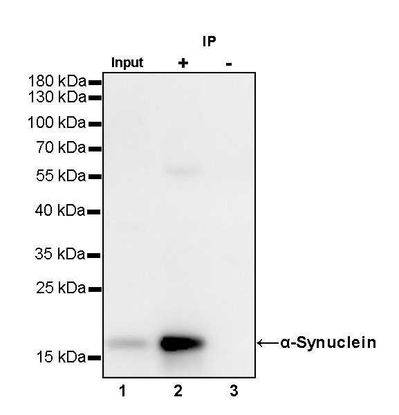
α-Synuclein Rabbit mAb at 1/50 dilution (1 ug) immunoprecipitating α-Synuclein in 0.4 mg mouse brain lysate. Western blot was performed on the immunoprecipitate using α-Synuclein Rabbit mAb at 1/1000 dilution. Secondary antibody (HRP) for IP was used at 1/400 dilution. Lane 1: mouse brain lysate 20 ug (Input) Lane 2: α-Synuclein Rabbit mAb IP in mouse brain lysate Lane 3: Rabbit monoclonal IgG IP in mouse brain lysate Predicted MW: 14 kDa Observed MW: 18 kDa Exposure time: 90 s