


 全部商品分类


Rabbit anti-β-Tubulin Monoclonal Antibody

 下载产品说明书
 用小程序,查商品更便捷


 收藏
收藏
 对比
对比 咨询
咨询
PBS, 40% Glycerol, 0.05%BSA, 0.03% Proclin 300
参考图片
WB result of β-Tubulin Rabbit mAb Primary antibody: β-Tubulin Rabbit mAb at 1/1000 dilution Lane 1: Hela whole cell lysate 20 µg Secondary antibody: #abs20040 at 1/10000 dilution Predicted MW: 55 kDa Observed MW: 55 kDa Exposure time: 15 seconds
WB result of β-Tubulin Rabbit mAb Primary antibody: β-Tubulin Rabbit mAb at 1/1000 dilution Lane 1:Mouse brain lysate 20 µg Lane 2: NIH/3T3 whole cell lysate 20 µg Secondary antibody: #abs20040 at 1/10000 dilution Predicted MW: 55 kDa Observed MW: 55 kDa Exposure time: Lane 1: 6 seconds; Lane 2: 15s
IHC shows positive staining in paraffin-embedded human testis. Anti-β-tubulin antibody was used at 1/1000 dilution, Secondary antibody: #abs20040 Counterstained with hematoxylin. Heat mediated antigen retrieval with Tris/EDTA buffer pH9.0 was performed before commencing with IHC staining protocol.
IHC shows positive staining in paraffin-embedded human cerebral cortex. Anti-β-tubulin antibody was used at 1/1000 dilution, Secondary antibody: #abs20040 Counterstained with hematoxylin. Heat mediated antigen retrieval with Tris/EDTA buffer pH9.0 was performed before commencing with IHC staining protocol.
IHC shows positive staining in paraffin-embedded mouse liver. Anti-β-tubulin antibody was used at 1/1000 dilution, Secondary antibody: #abs20040 Counterstained with hematoxylin. Heat mediated antigen retrieval with Tris/EDTA buffer pH9.0 was performed before commencing with IHC staining protocol.
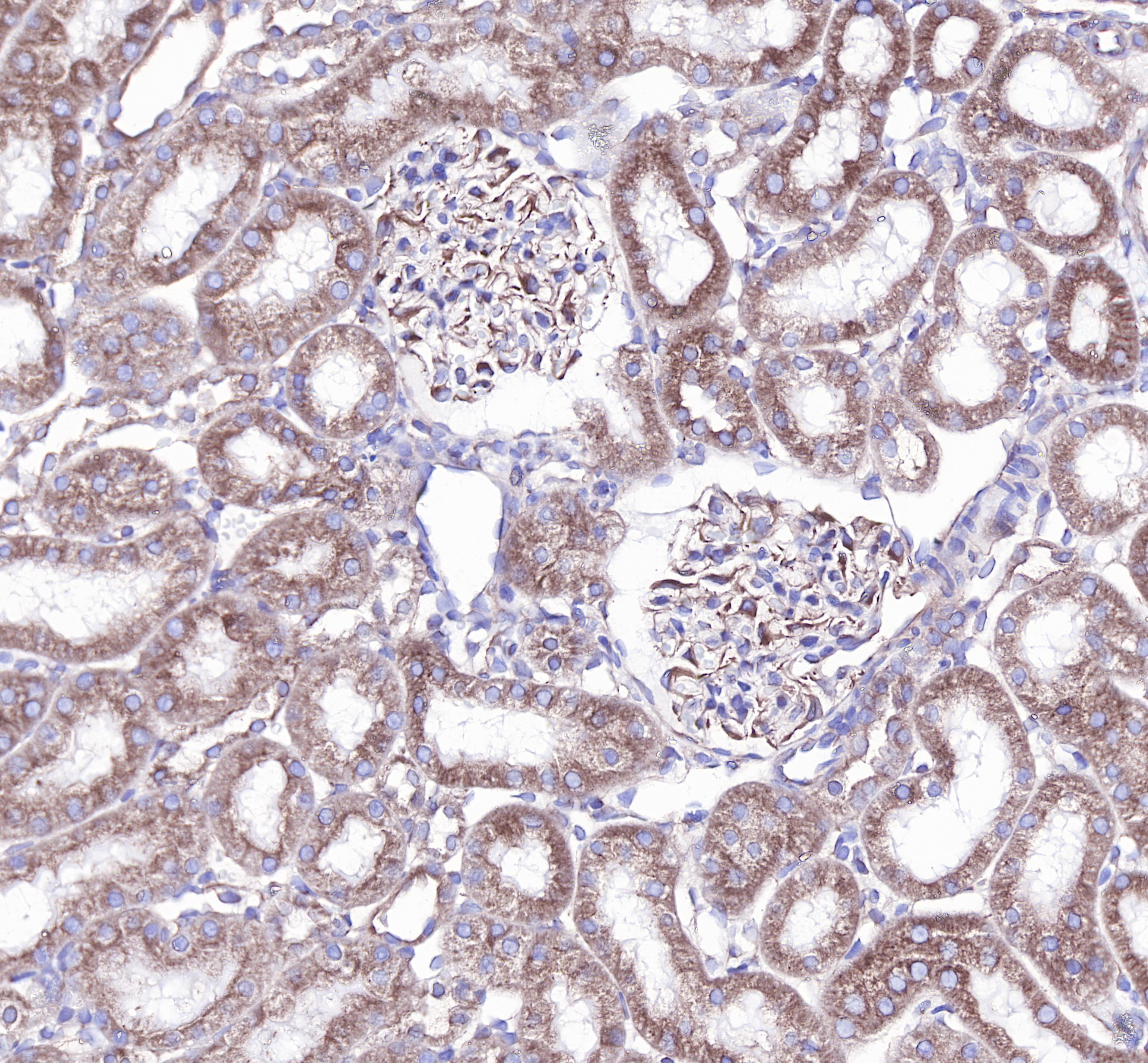
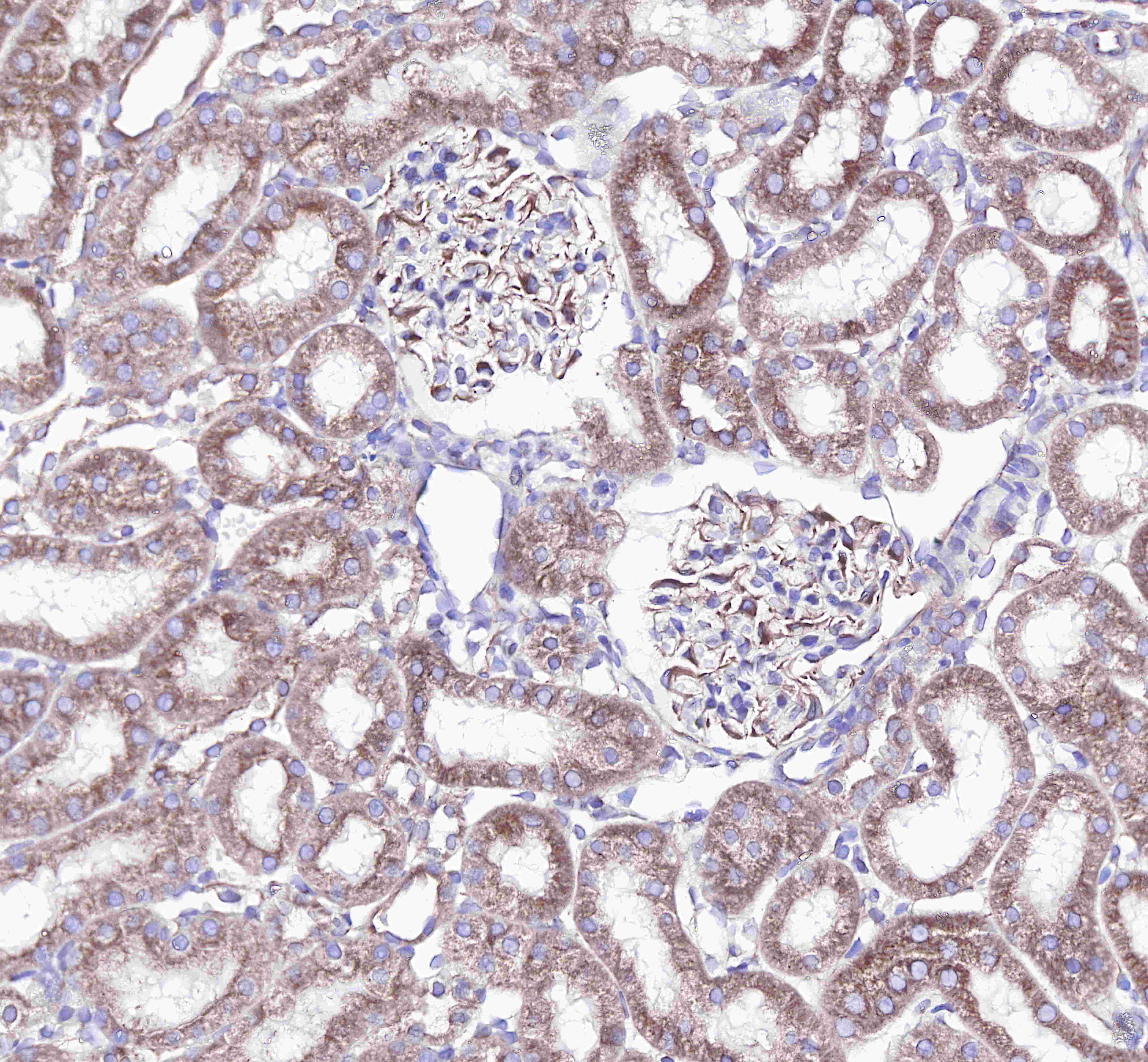
IHC shows positive staining in paraffin-embedded rat kidney. Anti-β-tubulin antibody was used at 1/1000 dilution, Secondary antibody: #abs20040 Counterstained with hematoxylin. Heat mediated antigen retrieval with Tris/EDTA buffer pH9.0 was performed before commencing with IHC staining protocol.
ICC shows cytoplasm staining in HeLa cells. Anti-β-tubulin antibody was used at 1/250 dilution and incubated overnight at 4°C. Secondary antibody: #abs20025 at 1/1000 dilution.The cells were fixed with 100% Methanol and permeabilized with 0.1% PBS-Triton X-100. Nuclei were countersained with DAPI.
ICC shows cytoplasm staining in NIH3T3 cells. Anti-β-tubulin antibody was used at 1/250 dilution and incubated overnight at 4°C. Secondary antibody: #abs20025 at 1/1000 dilution.The cells were fixed with 100% Methanol and permeabilized with 0.1% PBS-Triton X-100. Nuclei were countersained with DAPI.
Flow cytometric analysis of HeLa cells labelling β-tubulin antibody at 1/500 (0.1ug) dilution/ (red) compared with a Rabbit monoclonal IgG (Black) isotype control and an unlabelled control (cells without incubation with primary antibody and secondary antibody) (Blue). Secondary antibody:#abs20025.
IHC shows positive staining in paraffin-embedded human testis.
Anti-β-tubuLin antibody was used at 1/1000 dilution, followed by a Goat Anti-Rabbit IgG H&L (HRP) ready to use.
Counterstained with hematoxylin.
Heat mediated antigen retrieval with Tris/EDTA buffer pH9.0 was performed before commencing with IHC staining protocol.
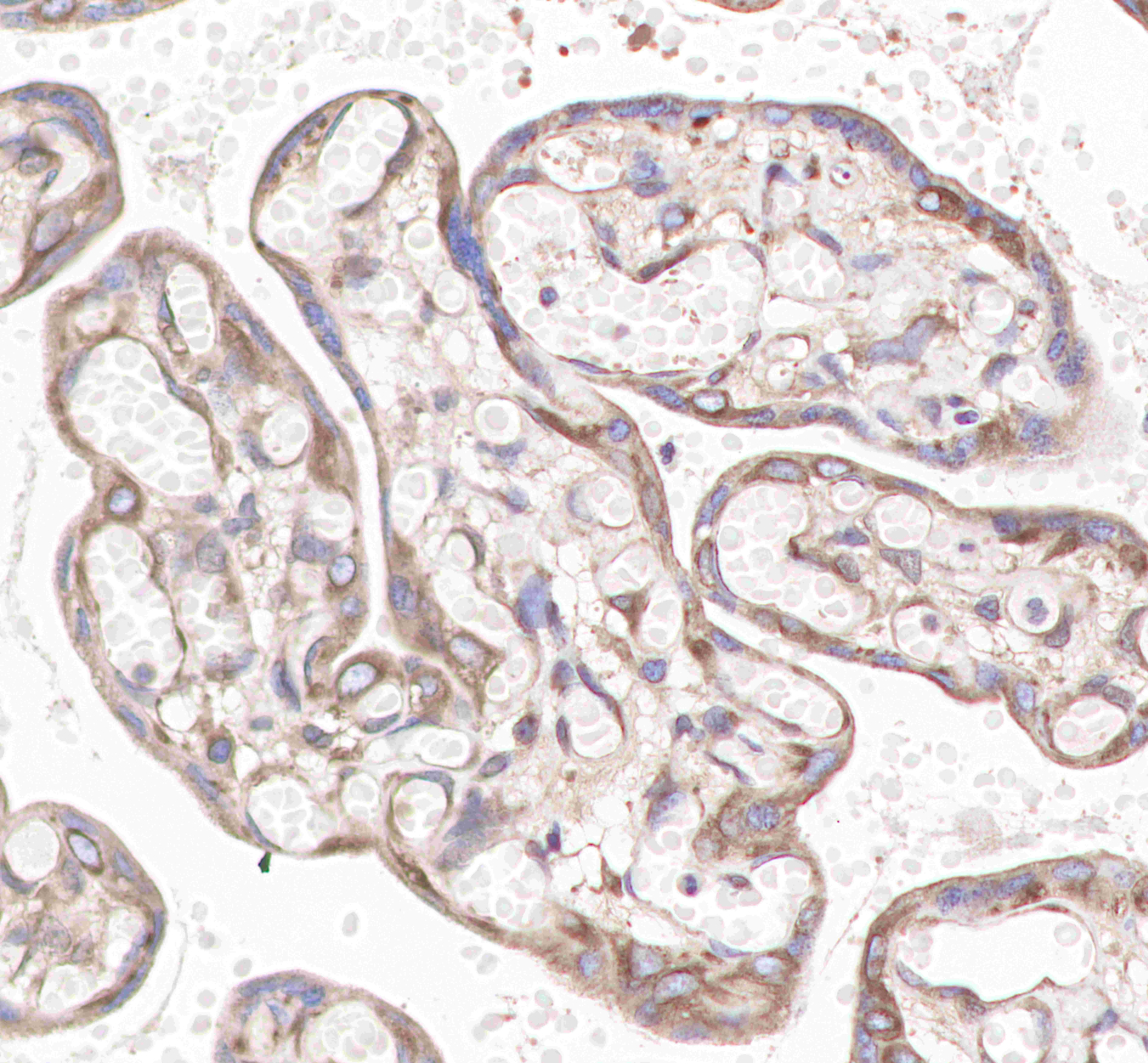
IHC shows positive staining in paraffin-embedded human placenta.
Anti-β-tubuLin antibody was used at 1/1000 dilution, followed by a Goat Anti-Rabbit IgG H&L (HRP) ready to use.
Counterstained with hematoxylin.
Heat mediated antigen retrieval with Tris/EDTA buffer pH9.0 was performed before commencing with IHC staining protocol.
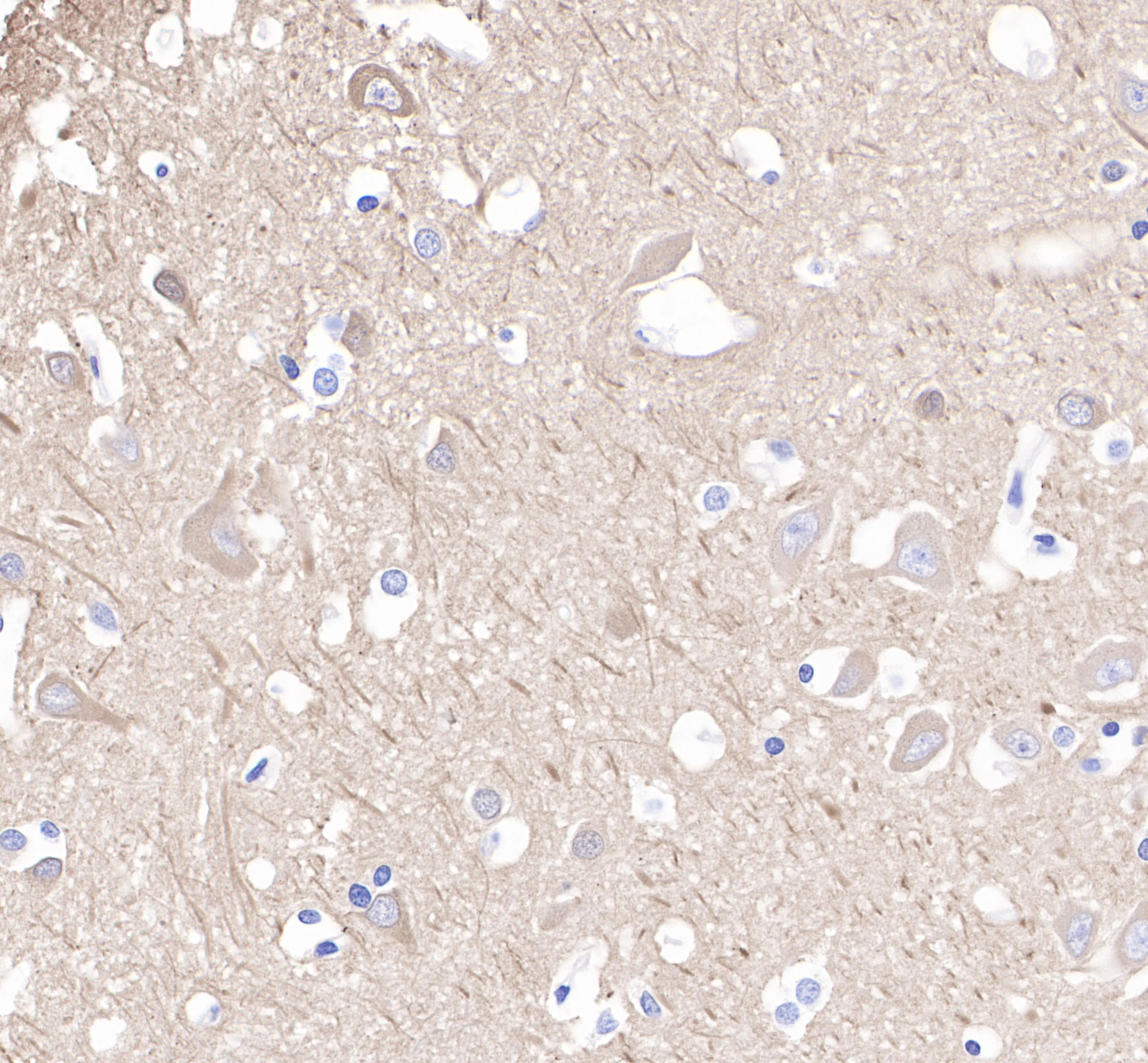
IHC shows positive staining in paraffin-embedded human cerebral cortex.
Anti-β-tubuLin antibody was used at 1/1000 dilution, followed by a Goat Anti-Rabbit IgG H&L (HRP) ready to use.
Counterstained with hematoxylin.
Heat mediated antigen retrieval with Tris/EDTA buffer pH9.0 was performed before commencing with IHC staining protocol.
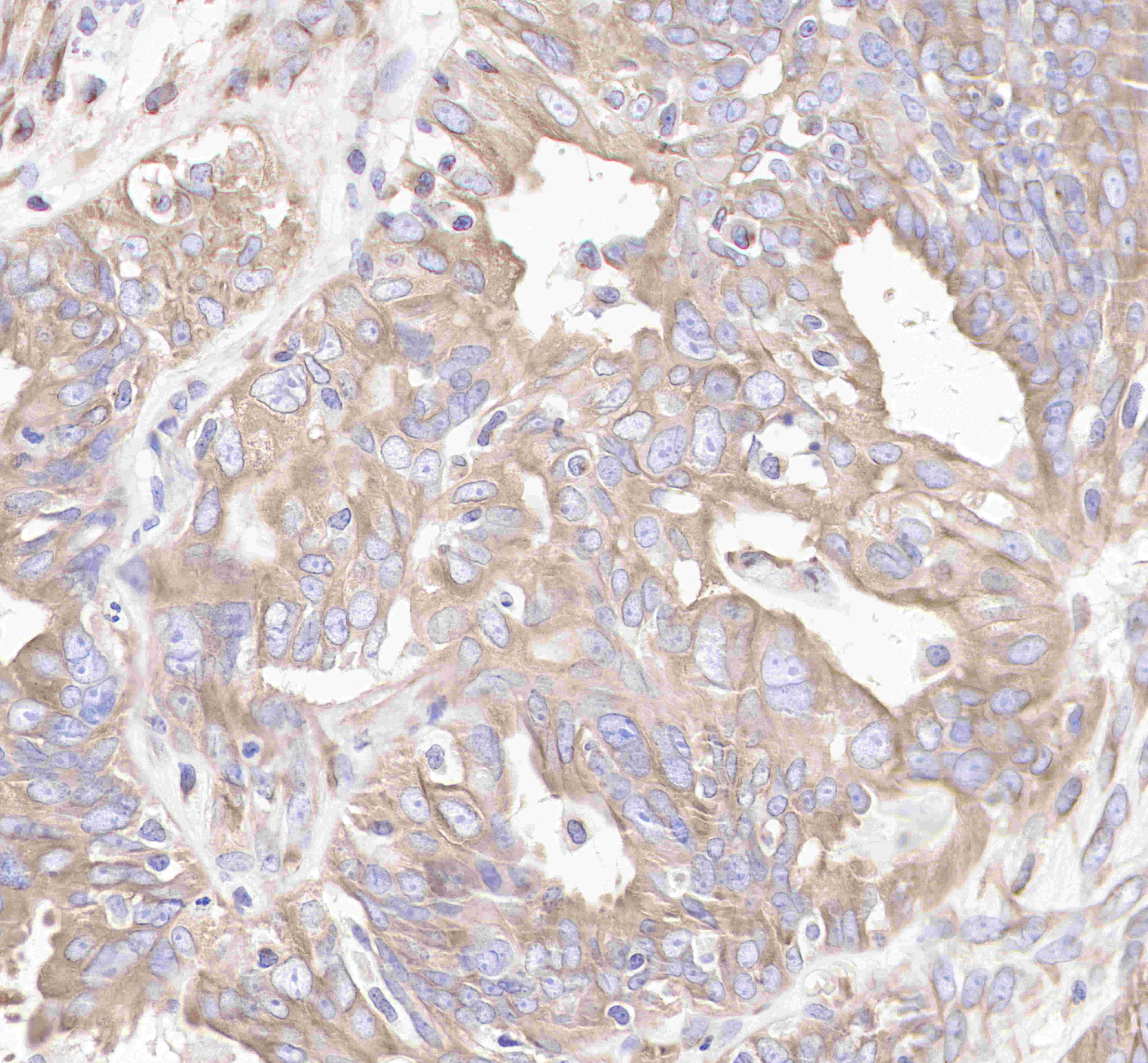
IHC shows positive staining in paraffin-embedded human colon cancer.
Anti-β-tubuLin antibody was used at 1/1000 dilution, followed by a Goat Anti-Rabbit IgG H&L (HRP) ready to use.
Counterstained with hematoxylin.
Heat mediated antigen retrieval with Tris/EDTA buffer pH9.0 was performed before commencing with IHC staining protocol.
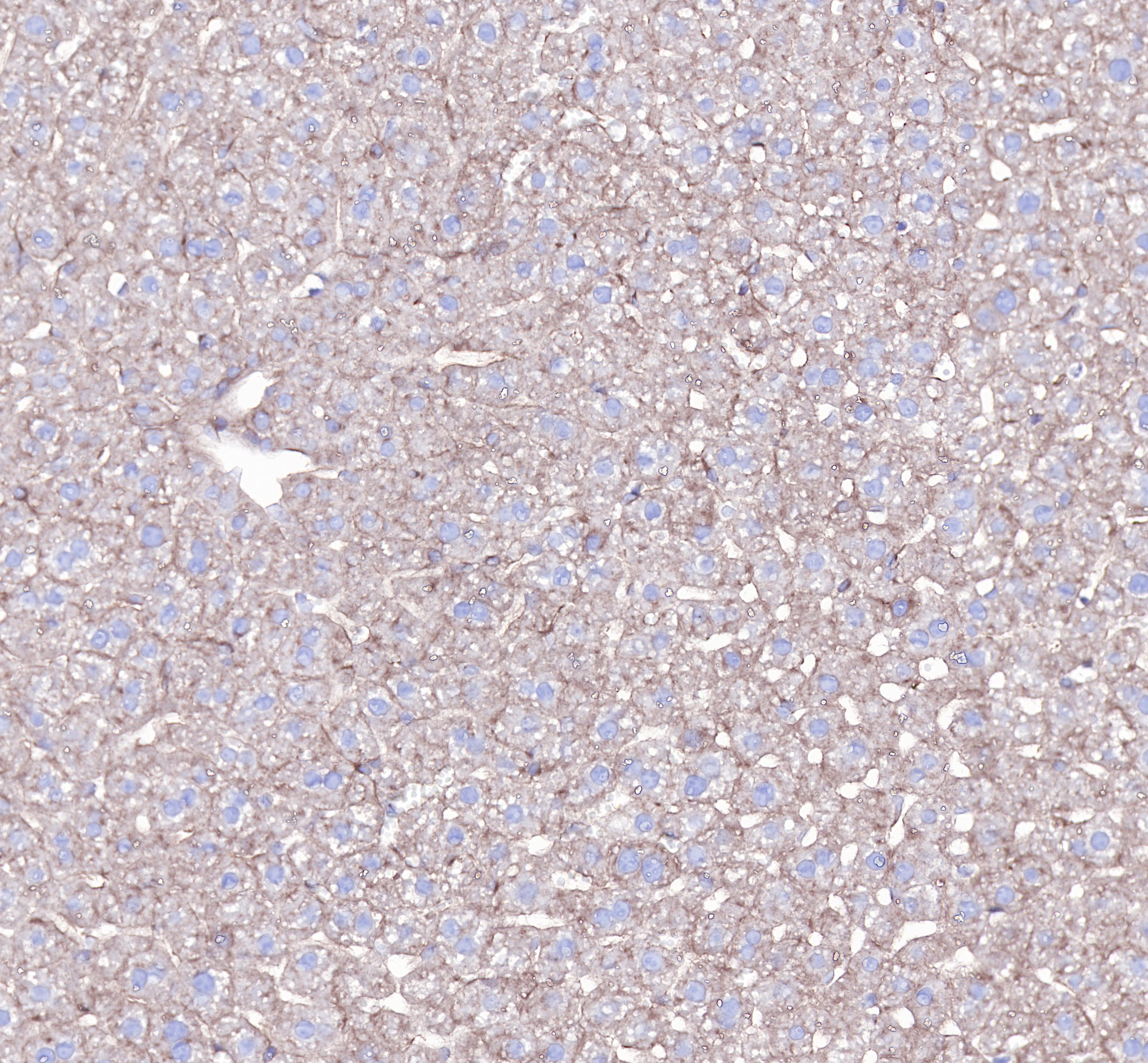
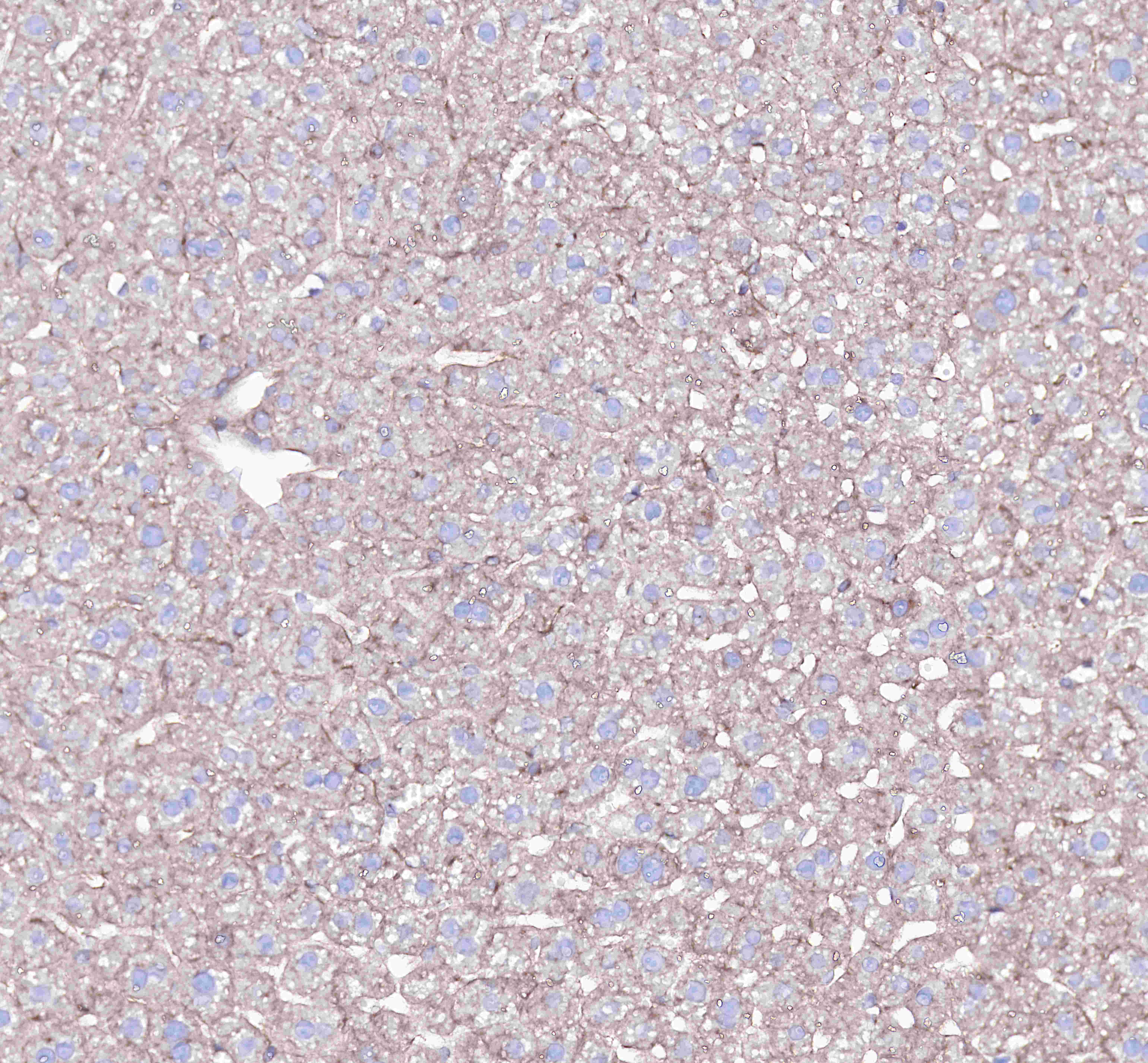
IHC shows positive staining in paraffin-embedded mouse liver.
Anti-β-tubuLin antibody was used at 1/1000 dilution, followed by a Goat Anti-Rabbit IgG H&L (HRP) ready to use.
Counterstained with hematoxylin.
Heat mediated antigen retrieval with Tris/EDTA buffer pH9.0 was performed before commencing with IHC staining protocol.
IHC shows positive staining in paraffin-embedded rat kidney.
Anti-β-tubuLin antibody was used at 1/1000 dilution, followed by a Goat Anti-Rabbit IgG H&L (HRP) ready to use.
Counterstained with hematoxylin.
Heat mediated antigen retrieval with Tris/EDTA buffer pH9.0 was performed before commencing with IHC staining protocol.
WB resuLt of β-TubuLin Rabbit mAb
Primary antibody: β-TubuLin Rabbit mAb at 1/1000 dilution
Lane 1: Hela whole cell lysate 20 µg
Secondary antibody: Goat Anti-Rabbit IgG, (H+L), HRP conjugated at 1/10000 dilution
Predicted MW: 50 kDa
Observed MW: 52 kDa
econds
WB resuLt of β-TubuLin Rabbit mAb
Primary antibody: β-TubuLin Rabbit mAb at 1/1000 dilution
Lane 1:Mouse brain lysate 20 µg
Lane 2: NIH/3T3 whole cell lysate 20 µg
Secondary antibody: Goat Anti-Rabbit IgG, (H+L), HRP conjugated at 1/10000 dilution
Predicted MW: 50 kDa
Observed MW: 52 kDa
WB resuLt of β-TubuLin Rabbit mAb
Primary antibody: β-TubuLin Rabbit mAb at 1/1000 dilution
Lane 1:rat brain lysate 20 µg
Secondary antibody: Goat Anti-Rabbit IgG, (H+L), HRP conjugated at 1/10000 dilution
Predicted MW: 50 kDa
Observed MW: 52 kDa
Flow cytometric analysis of HeLa cells labelling β-tubuLin antibody at 1/500 (0.1ug) dilution/ (red) compared with a Rabbit monoclonal IgG (Black) isotype control and an unlabelled control (cells without incubation with primary antibody and secondary antibody) (Blue). Goat Anti-Rabbit IgG Alexa Fluor® 488 was used as the secondary antibody.
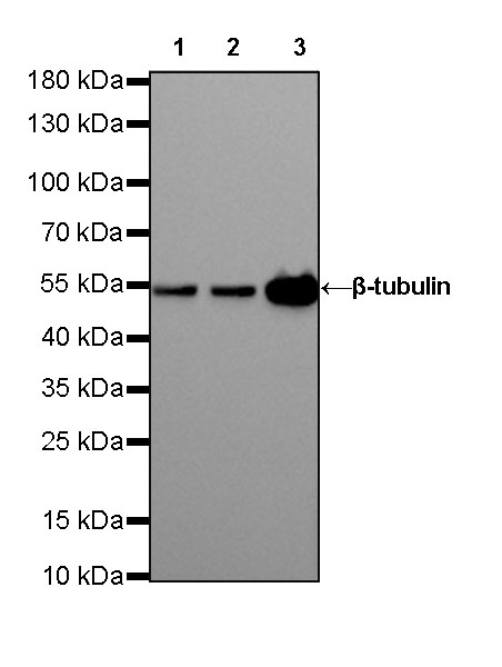
WB resuLt of β-TubuLin Rabbit mAb
Primary antibody: β-TubuLin Rabbit mAb at 1/10000 dilution
Lane 1: HeLa whole cell lysate 20 µg
Lane 2: NIH/3T3 whole cell lysate 20 µg
Lane 3: rat brain lysate 20 µg
Secondary antibody: Goat Anti-Rabbit IgG, (H+L), HRP conjugated at 1/10000 dilution
Predicted MW: 50 kDa
Observed MW: 52 kDa
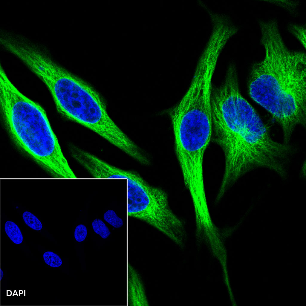
ICC shows positive staining in HeLa cells. Anti-β-tubuLin antibody was used at 1/500 dilution (Green) and incubated overnight at 4°C. Goat polyclonal Antibody to Rabbit IgG - H&L (Alexa Fluor® 488) was used as secondary antibody at 1/1000 dilution. The cells were fixed with 4% PFA and permeabilized with 0.1% PBS-Triton X-100. Nuclei were counterstained with DAPI (Blue).