


 全部商品分类
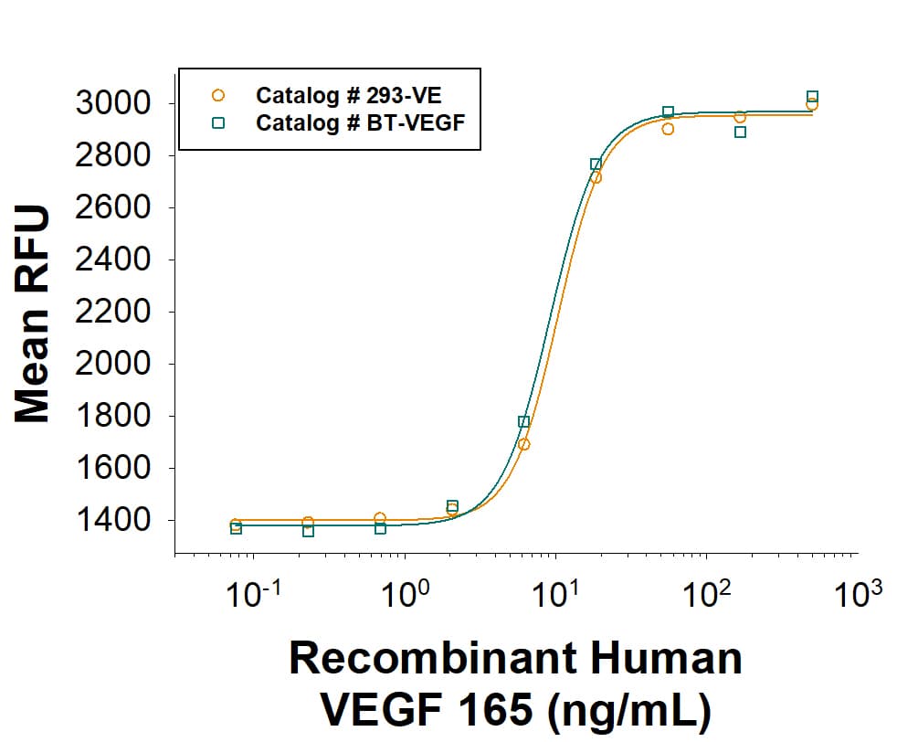


 下载产品说明书 下载SDS
 用小程序,查商品更便捷


 收藏
收藏
 对比
对比 咨询
咨询Scientific Data
 View Larger
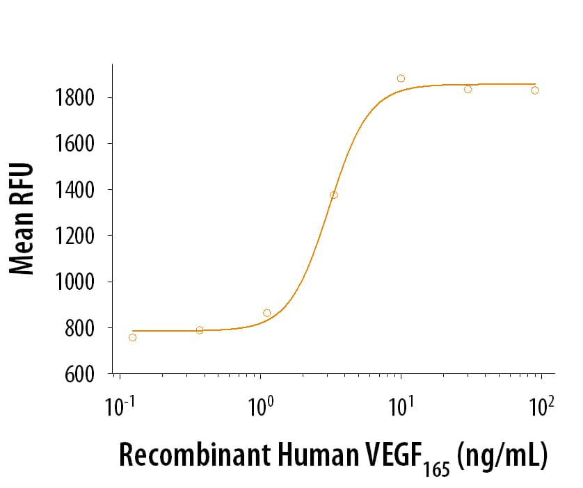
Recombinant Human VEGF165(Catalog # 293-VE) stimulates proliferation in HUVEC human umbilical vein endothelial cells. The ED50 is 1-6 ng/mL.
 View Larger
As an alternative, please consider our next generation Recombinant Human VEGF 165 (Catalog # BT-VEGF). It has equivalent bioactivity to Recombinant Human VEGF 165 (Catalog # 293-VE). It combines R&D Systems quality with scalability that allows for a solid supply chain. Both Recombinant Human VEGF 165 proteins are measured in a cell proliferation assay using HUVEChuman umbilical vein endothelial cells.
 View Larger
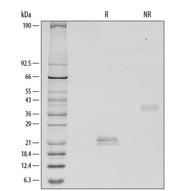
1 μg/lane of Recombinant Human VEGF165was resolved by SDS-PAGE with silver staining, under reducing (R) and non-reducing (NR) conditions, showing major bands at 20-22 kDa and 39-42 kDa, respectively. Multiple bands in gel are due to glycosylation.
Carrier Free
CF stands for Carrier Free (CF). We typically add Bovine Serum Albumin (BSA) as a carrier protein to our recombinant proteins. Adding a carrier protein enhances protein stability, increases shelf-life, and allows the recombinant protein to be stored at a more dilute concentration. The carrier free version does not contain BSA.
In general, we advise purchasing the recombinant protein with BSA for use in cell or tissue culture, or as an ELISA standard. In contrast, the carrier free protein is recommended for applications, in which the presence of BSA could interfere.
293-VE
| Formulation | Lyophilized from a 0.2 μm filtered solution in Acetonitrile and TFA with BSA as a carrier protein. |
| Reconstitution | Reconstitute at 100 μg/mL in sterile PBS containing at least 0.1% human or bovine serum albumin. Alternatively, reconstitute at 100‑500 μg/mL in sterile 4 mM HCl containing 0.1% human or bovine serum albumin. |
| Shipping | The product is shipped at ambient temperature. Upon receipt, store it immediately at the temperature recommended below. |
| Stability & Storage: | Use a manual defrost freezer and avoid repeated freeze-thaw cycles.
|
293-VE/CF
| Formulation | Lyophilized from a 0.2 μm filtered solution in Acetonitrile and TFA with trehalose. |
| Reconstitution | Reconstitute at 100 μg/mL in sterile PBS. Alternatively, reconstitute at 100-500 μg/mL in sterile 4 mM HCl. |
| Shipping | The product is shipped at ambient temperature. Upon receipt, store it immediately at the temperature recommended below. |
| Stability & Storage: | Use a manual defrost freezer and avoid repeated freeze-thaw cycles.
|
Recombinant Human VEGF 165 Protein Summary
Product Specifications
Ala27-Arg191
Analysis
39-42 kDa, under non-reducing conditions.
Background: VEGF
Vascular endothelial growth factor (VEGF or VEGF-A), also known as vascular permeability factor (VPF), is a potent mediator of both angiogenesis and vasculogenesis in the fetus and adult (1-3). It is a member of the PDGF family that is characterized by the presence of eight conserved cysteine residues and a cystine knot structure (4). Humans express alternately spliced isoforms of 121, 145, 165, 183, 189, and 206 amino acids (aa) in length (4). VEGF165 appears to be the most abundant and potent isoform, followed by VEGF121 and VEGF189 (3, 4). Isoforms other than VEGF121 contain basic heparin-binding regions and are not freely diffusible (4). Human VEGF165 shares 88% aa sequence identity with corresponding regions of mouse and rat, 96% with porcine, 95% with canine, and 93% with feline, equine and bovine VEGF, respectively. VEGF binds the type I transmembrane receptor tyrosine kinases VEGF R1 (also called Flt-1) and VEGF R2 (Flk-1/KDR) on endothelial cells (4). Although VEGF affinity is highest for binding to VEGF R1, VEGF R2 appears to be the primary mediator of VEGF angiogenic activity (3, 4). VEGF165 binds the semaphorin receptor, Neuropilin-1 and promotes complex formation with VEGF R2 (5). VEGF is required during embryogenesis to regulate the proliferation, migration, and survival of endothelial cells (3, 4). In adults, VEGF functions mainly in wound healing and the female reproductive cycle (3). Pathologically, it is involved in tumor angiogenesis and vascular leakage (6, 7). Circulating VEGF levels correlate with disease activity in autoimmune diseases such as rheumatoid arthritis, multiple sclerosis and systemic lupus erythematosus (8). VEGF is induced by hypoxia and cytokines such as IL-1, IL-6, IL-8, oncostatin M and TNF-alpha (3, 4, 9).
Due to its role in angiogenesis of blood vessels, tumor and stroma cells use VEGF to stimulate formation of blood vessels and the proliferation and survival of endothelial cells. Specific immunotherapies targeting the VEGF signaling pathway include the recombinant antibody against VEGF (Bevacizumab), antibodies targeting the main VEGF receptor (VEGFR2), and small molecule inhibitors against VEGF receptor tyrosine kinases (10). Immune checkpoint inhibitors are an important tool in cancer therapies as tumor cells can hijack immune checkpoint signals to evade detection by immune cells. In addition to stimulating the formation of tumor blood vessels, VEGF has immunosuppressive effects by acting on dendritic cells to block their antigen-presenting and T cell stimulatory functions. Targeting VEGF in combination with other immune checkpoint ligands or receptors may prove more effective in immunotherapy approaches to certain cancer types (11). Because of its role in the formation of blood vessels, VEGF is also an important factor in skeletal development where blood supply and vascularization are crucial. This has made VEGF an important molecule in regenerative studies for bone repair as sustained release of VEGF has been shown to improve the efficiency of bone regeneration (12).
In differentiation protocols for stems cells, VEGF is a commonly added growth factor for the transformation of induced pluripotent stem cells into hematopoietic progenitor cells used to make Natural Killer cells (13, 14). VEGF has also been used to transform intermediate mesoderm into kidney glomerular podocytes or stem cell-derived liver spheres (15, 16). VEGF may also be used in assistance of stem cell transplantations by supporting angiogenesis at sites of stem cell transplants or as a honing tool for adipose-derived mesenchymal stem cells or bone marrow stem cells to migrate to (17, 18).- Leung, D.W. et al. (1989) Science 246:1306.
- Keck, P.J. et al. (1989) Science 246:1309.
- Byrne, A.M. et al. (2005) J. Cell. Mol. Med. 9:777.
- Robinson, C.J. and S.E. Stringer (2001) J. Cell. Sci. 114:853.
- Pan, Q. et al. (2007) J. Biol. Chem. 282:24049.
- Weis, S.M. and D.A. Cheresh (2005) Nature 437:497.
- Thurston, G. (2002) J. Anat. 200:575.
- Carvalho, J.F. et al. (2007) J. Clin. Immunol. 27:246.
- Angelo, L.S. and R. Kurzrock (2007) Clin. Cancer Res. 13:2825.
- Apte, R.S. et al. (2019) Cell 176(6):1248.
- Sangro, B. et al. (2021) Nature 18:525-543.
- Hu, K. & Olsen, B.R. (2016) Bone 91:30-38.
- Zhou, Y. et al. (2022) Cancers 14:2266.
- Li, Y. et al. (2018) Cell Stem Cell 23(2):181-192.
- Musah, S. et al. (2018) Nat. Protoc. 13(7):1662-1685.
- Meseguer-Ripolles, J. et al. (2021) STAR Protoc. 2(2):100502.
- Hutchings, G. et al. (2020) Int. J. Mol. Sci. 21(11):3790.
- Zhang, W. et al. (2014) Euro Cell and Mater. 27:1-12.

参考图片
1 μg/lane of Recombinant Human VEGF165 was resolved by SDS-PAGE with silver staining, under reducing (R) and non-reducing (NR) conditions, showing major bands at 20-22 kDa and 39-42 kDa, respectively. Multiple bands in gel are due to glycosylation.
Recombinant Human VEGF165 (Catalog # 293-VE) stimulates proliferation in HUVEC human umbilical vein endothelial cells. The ED50 is typically 1-6 ng/mL.