


 全部商品分类


1/3
BD Transduction Laboratories™ Purified Mouse Anti-RIP


品牌: BD Pharmingen
 下载产品说明书 下载SDS
 用小程序,查商品更便捷


 收藏
收藏
 对比
对比 咨询
咨询反应种属:
Human (QC Testing), Mouse, Rat, Dog, Chicken (Tested in Development)
来源宿主:
Mouse IgG2a
产品介绍
产品介绍
产品信息
抗原名称
RIP

宿主
Mouse IgG2a
免疫原
Human RIP aa. 385-650
简单描述
Binding or cross linking of the Fas antigen (also known as APO-1 and CD95) is known to elicit apoptosis in susceptible cells. Fas is a member of a family of cell surface receptors which includes tumor necrosis factor receptors (TNF-R, and TNF-R2) and nerve growth factor receptors (NGF-R), CD40, OX40, CD30, CD27, and 4-1BB. Several members of this family have been shown to regulate or induce cell death (TNF-R1 and TNF-R2). A 74 kDa member of this family protein named RIP (Receptor Interacting Protein) contains an N-terminal region with homology to protein kinases and a C-terminal region containing a cytoplasmic "death domain" present in both Fas and TNF-R1. Both Fas and RIP have been shown to require this death domain to induce apoptosis and overexpression of RIP has been shown to induce cell death in transfected cells.
商品描述
38/RIP
Binding or cross linking of the Fas antigen (also known as APO-1 and CD95) is known to elicit apoptosis in susceptible cells. Fas is a member of a family of cell surface receptors which includes tumor necrosis factor receptors (TNF-R, and TNF-R2) and nerve growth factor receptors (NGF-R), CD40, OX40, CD30, CD27, and 4-1BB. Several members of this family have been shown to regulate or induce cell death (TNF-R1 and TNF-R2). A 74 kDa member of this family protein named RIP (Receptor Interacting Protein) contains an N-terminal region with homology to protein kinases and a C-terminal region containing a cytoplasmic "death domain" present in both Fas and TNF-R1. Both Fas and RIP have been shown to require this death domain to induce apoptosis and overexpression of RIP has been shown to induce cell death in transfected cells.
同种型
Mouse IgG2a
克隆号
克隆 38/RIP (RUO)
浓度
250 µg/ml
产品详情
Purified
Tissue culture supernatant is purified by either protein A/G or affinity purification methods. Both methods yield antibody in solution that is free of most other soluble proteins, lipids, etc. This format provides pure antibody that is suitable for a number of downstream applications including: secondary labeling for flow cytometry or microscopy, ELISA, Western blot, etc.
应用
实验应用
Western blot (Routinely Tested), Immunofluorescence, Immunoprecipitation (Tested During Development), Immunohistochemistry (Not Recommended)
反应种属
Human (QC Testing), Mouse, Rat, Dog, Chicken (Tested in Development)
目标/特异性
RIP
背景
别名
Receptor Interacting Protein
制备和贮存
存储溶液
Aqueous buffered solution containing BSA, glycerol, and ≤0.09% sodium azide.
保存方式
Aqueous buffered solution containing BSA, glycerol, and ≤0.09% sodium azide.
文献
文献
研发参考(5)
1. Devin A, Lin Y, Yamaoka S, Li Z, Karin M, Liu Zg. The alpha and beta subunits of IkappaB kinase (IKK) mediate TRAF2-dependent IKK recruitment to tumor necrosis factor (TNF) receptor 1 in response to TNF. Mol Cell Biol. 2002; 21(12):3986-3994. (Biology: Western blot).
2. Fulda S, Meyer E, Debatin KM. Metabolic inhibitors sensitize for CD95 (APO-1/Fas)-induced apoptosis by down-regulating Fas-associated death domain-like interleukin 1-converting enzyme inhibitory protein expression. Cancer Res. 2000; 60(14):3947-3956. (Biology: Western blot).
3. Lewis J, Devin A, Miller A, et al. Disruption of hsp90 function results in degradation of the death domain kinase, receptor-interacting protein (RIP), and blockage of tumor necrosis factor-induced nuclear factor-kappaB activation. J Biol Chem. 2000; 275(14):10519-10526. (Biology: Immunoprecipitation, Western blot).
4. Stanger BZ, Leder P, Lee TH, Kim E, Seed B. RIP: a novel protein containing a death domain that interacts with Fas/APO-1 (CD95) in yeast and causes cell death. Cell. 1995; 81(4):513-523. (Biology).
5. Takahashi T, Tanaka M, Brannan CI, et al. Generalized lymphoproliferative disease in mice, caused by a point mutation in the Fas ligand. Cell. 1994; 76(6):969-976. (Biology).
参考图片
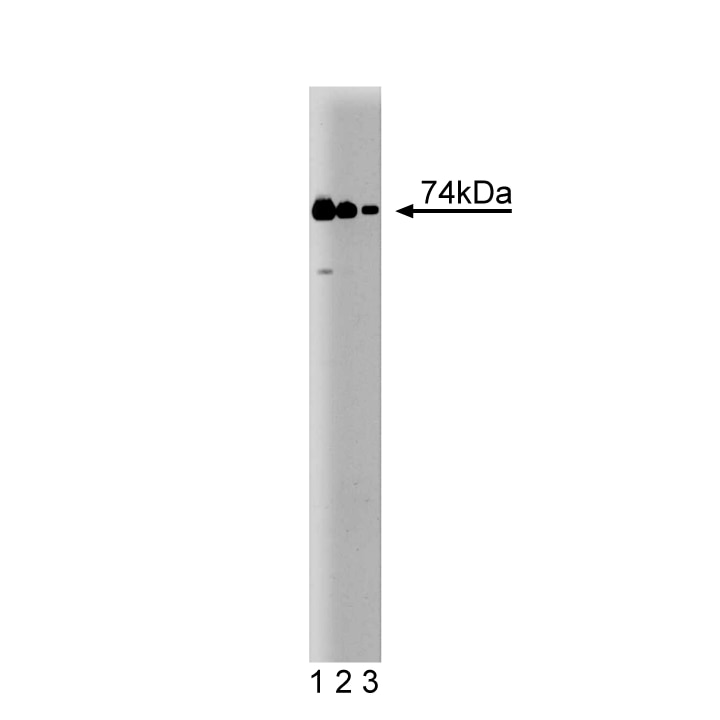
Western blot analysis of RIP on a human endothelial cell lysate. Lane 1: 1:1000, lane 2: 1:2000, lane 3: 1:4000 dilution of the mouse anti-RIP antibody.
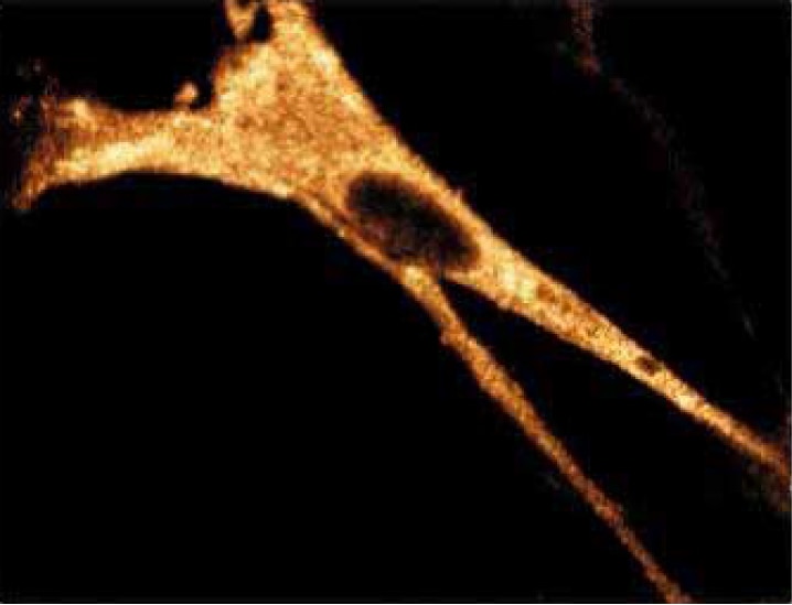
Immunofluorescence staining of WI-38 cells (Human lung fibroblasts; ATCC CCL-75).
Western blot analysis of RIP on a human endothelial cell lysate. Lane 1: 1:1000, lane 2: 1:2000, lane 3: 1:4000 dilution of the mouse anti-RIP antibody.
Immunofluorescence staining of WI-38 cells (Human lung fibroblasts; ATCC CCL-75).
声明 :本官网所有报价均为常温或者蓝冰运输价格,如有产品需要干冰运输,需另外加收干冰运输费。