


 全部商品分类
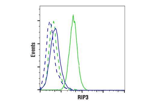


 用小程序,查商品更便捷


Monoclonal antibody is produced by immunizing animals with a synthetic peptide corresponding to residues near the carboxy terminus of human RIP3 protein.
Product Usage Information
| Application | Dilution |
|---|---|
| Western Blotting | 1:1000 |
| Immunoprecipitation | 1:50 |
| Immunohistochemistry (Paraffin) | 1:50 - 1:200 |
| Immunofluorescence (Immunocytochemistry) | 1:100 - 1:400 |
| Flow Cytometry (Fixed/Permeabilized) | 1:400 - 1:1600 |
Specificity/Sensitivity
Species Reactivity:
Human
Supplied in 10 mM sodium HEPES (pH 7.5), 150 mM NaCl, 100 µg/ml BSA, 50% glycerol and less than 0.02% sodium azide. Store at –20°C. Do not aliquot the antibody.
参考图片
Flow cytometric analysis of SU-DHL-4 cells (blue, negative) and THP-1 cells (green, positive) using RIP3 (E7A7F) XP® Rabbit mAb (solid lines) or a concentration-matched Rabbit (DA1E) mAb IgG XP® Isotype Control #3900 (dashed lines). Anti-rabbit IgG (H+L), F(ab')2 Fragment (Alexa Fluor® 488 Conjugate) #4412 was used as a secondary antibody.
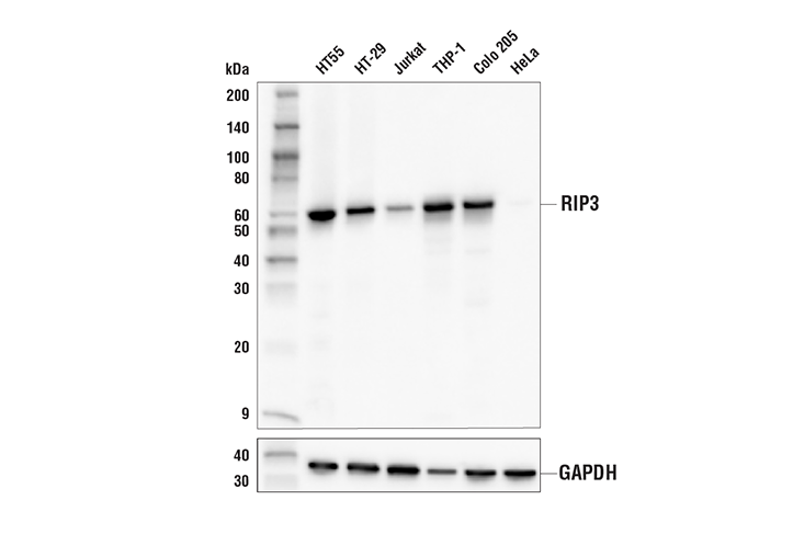
Western blot analysis of extracts from various cell lines using RIP3 (E7A7F) XP® Rabbit mAb (upper) or GAPDH (D16H11) XP® Rabbit mAb #5174 (lower). Absence of signal in HeLa cells is predicted from published reports of epigenetic loss of expression and confirms the specificity of the antibody for RIP3.
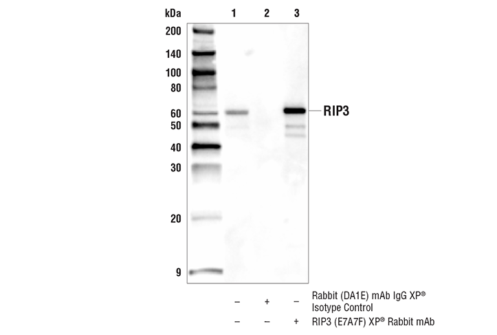
Immunoprecipitation of RIP3 protein from THP-1 cell extracts. Lane 1 is 10% input, lane 2 is Rabbit (DA1E) mAb IgG XP® Isotype Control #3900, and lane 3 is RIP3 (E7A7F) XP® Rabbit mAb. Western blot analysis was performed using RIP3 (E7A7F) XP® Rabbit mAb. Mouse Anti-rabbit IgG (Conformation Specific) (L27A9) mAb (HRP Conjugate) #5127 was used as a secondary antibody.
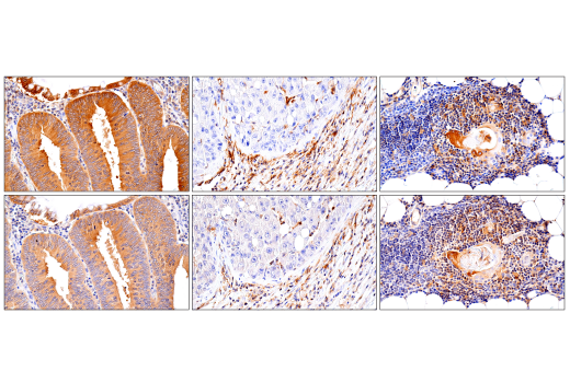
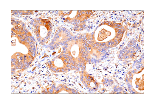
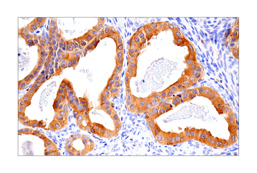
Immunohistochemical analysis of paraffin-embedded human colon carcinoma (left), hepatocellular carcinoma (middle), or normal thymus (right) using RIP3 (E7A7F) XP® Rabbit mAb (top) or RIP3 Rabbit mAb (bottom). These two antibodies detect independent, unique epitopes on human RIP3 protein. The similar staining patterns obtained with both antibodies help to confirm the specificity of the staining.
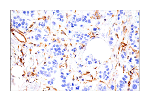
Immunohistochemical analysis of paraffin-embedded human ductal breast carcinoma using RIP3 (E7A7F) XP® Rabbit mAb.
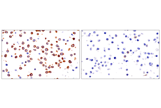
Immunohistochemical analysis of paraffin-embedded HT-29 cell pellet (left, high-expressing) or HeLa cell pellet (right, low-expressing) using RIP3 (E7A7F) XP® Rabbit mAb.
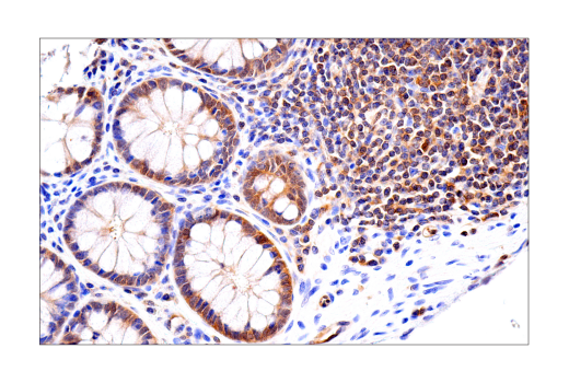
Immunohistochemical analysis of paraffin-embedded normal human colon using RIP3 (E7A7F) XP® Rabbit mAb.
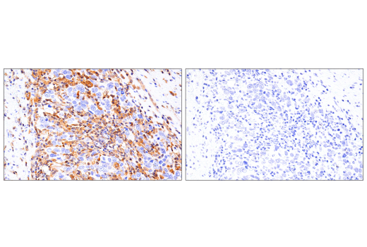
Immunohistochemical analysis of paraffin-embedded human colon carcinoma using RIP3 (E7A7F) XP® Rabbit mAb (left) compared to concentration-matched Rabbit (DA1E) mAb IgG XP® Isotype Control #3900 (right).
Immunohistochemical analysis of paraffin-embedded human esophageal adenocarcinoma using RIP3 (E7A7F) XP® Rabbit mAb.
Immunohistochemical analysis of paraffin-embedded human squamous cell carcinoma of the tonsil using RIP3 (E7A7F) XP® Rabbit mAb.
Immunohistochemical analysis of paraffin-embedded human endometrioid adenocarcinoma using RIP3 (E7A7F) XP® Rabbit mAb.
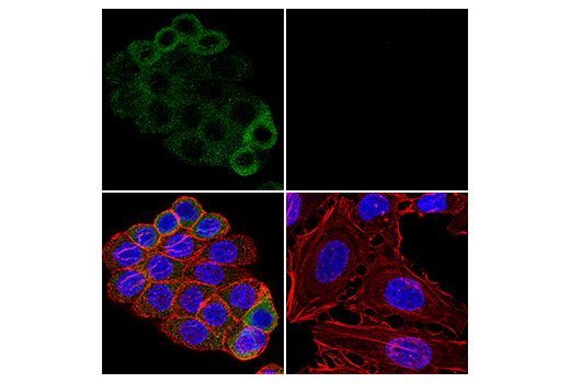
Confocal immunofluorescent analysis of HT-29 cells (left, positive) or HeLa cells (right, negative) using RIP3 (E7A7F) XP® Rabbit mAb (green). Actin filaments were labeled with DyLight™ 554 Phalloidin #13054 (red). Samples were mounted in ProLong® Gold Antifade Reagent with DAPI #8961 (blue).