


 全部商品分类
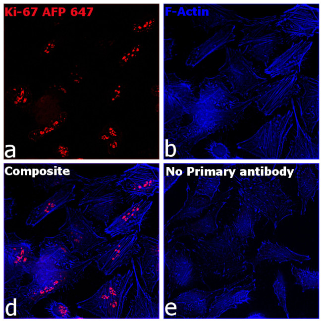


RT KI67 AFP647 RMAB

 下载产品说明书 下载COA 下载SDS
 用小程序,查商品更便捷


 收藏
收藏
 对比
对比 咨询
咨询种属反应
宿主/亚型
Expression System
分类
类型
克隆号
偶联物
激发/发射光谱

形式
浓度
纯化类型
保存液
内含物
保存条件
运输条件
RRID
产品详细信息
Alexa Fluor™ Plus recombinant antibodies are conjugated using new, proprietary dye chemistry so you can generate stunning data. Alexa Fluor™ Plus antibodies represent an advancement in fluorescent conjugate technology. Alexa Fluor™ Plus antibodies provide brighter signal compared to leading Alexa Fluor™ antibodies, providing you with better signal-to-noise for your critical experiments. These antibodies show better specificity and lot-to-lot consistency as these are recombinant antibodies, generated by cloning specific genes for the desired antibodies into an expression vector and expressed in vitro.
Using conjugate solutions: Centrifuge the protein conjugate solution briefly in a microcentrifuge before use; add only the supernatant to the experiment. This step will help eliminate any protein aggregates that may have formed during storage, thereby reducing nonspecific background staining.
Applications Tested: This SolA15 antibody has been tested by immunohistochemistry of mouse spleen tissue and immunocytochemistry and flow cytometric analysis of HeLa cells. This may be used for immunocytochemistry at 0.5 µg/mL and for flow cytometry at less than or equal to 0.02-0.25 µg per test. A test is defined as the amount (µg) of antibody that will stain a cell sample in a final volume of 100 µL. Cell number should be determined empirically but can range from 10^5 to 10^8 cells/test. It is recommended that the antibody be carefully titrated for optimal performance in the assay of interest.
Excitation: 658 nm; Emission: 675 nm; Laser: Red Laser
Filtration: 0.2 µm post-manufacturing filtered.
靶标信息
Ki-67 is a nuclear protein that is expressed during various stages in the cell cycle, particularly during late G1, S, G2, and M phases. The protein has a forkhead associated domain (FHA) through which it associates with euchromatin at the perichromosomal layer, the centromeric heterochromatin, and the nucleolus. Ki-67 is shown to have a cell cycle dependent topographical distribution with perinucleolar expression at G1, expression in the nuclear matrix at G2, and expression on the chromosomes during M phase. Ki-67 is commonly used as a proliferation marker because it is not detected in G0 cells, but increases steadily from G1 through mitosis. Ki-67 antibodies are useful in establishing the cell growing fraction in neoplasms. In neoplastic tissues, the prognostic value is comparable to the tritiated thymidine-labelling index. The correlation between low Ki-67 index and histologically low-grade tumors is strong. Ki-67 is routinely used as a neuronal marker of cell cycling and proliferation.
仅用于科研。不用于诊断过程。未经明确授权不得转售。
生物信息学
蛋白别名: Antigen identified by monoclonal antibody Ki-67; Antigen identified by monoclonal antibody Ki-67 homolog; Antigen KI-67; Antigen KI-67 homolog; Proliferation marker protein Ki-67; proliferation-related Ki-67 antigen; protein phosphatase 1, regulatory subunit 105; RP11-380J17.2
基因别名: D630048A14Rik; Ki-67; Ki67; KIA; MIB-; MIB-1; MKI67; PPP1R105
UniProt ID:(Human) P46013, (Mouse) E9PVX6
Entrez Gene ID:(Human) 4288, (Mouse) 17345
参考图片
Immunofluorescence analysis of Ki-67 was performed using 70% confluent HeLa cells. The cells were fixed with 4% paraformaldehyde for 10 minutes, permeabilized with 0.5% Triton X-100 for 10 minutes, and blocked with 2% BSA for 1 hour at room temperature. The cells were stained with Ki-67 Recombinant Rat Monoclonal Antibody (SolA15), Alexa Fluor™ Plus 647 (Product # 740008TP647,1:200) at 4 degree Celsius overnight. Panel a) shows representative images of cells that were stained for detection and localization of Ki-67. Panel b) represents cytoskeletal F-actin staining using Alexa Fluor™ Plus 405 Phalloidin (Product # A30104, 1:300). Panel c) is a composite image of panels a, and b, clearly demonstrating nuclear localization of Ki-67. Panel d) represents no primary antibody control to assess the background. The images were captured at 60X magnification.
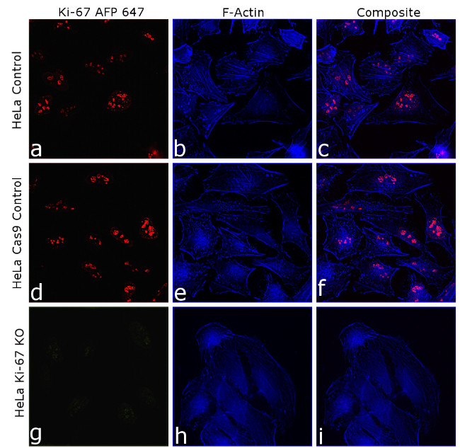
Knockout of Ki-67 was achieved by CRISPR-Cas9 genome editing using LentiArray™ Lentiviral sgRNA (Product # A32042, Assay ID CRISPR955686_LV) and LentiArray Cas9 Lentivirus (Product # A32064). Immunofluorescence analysis was performed on HeLa wild type cells (panel a-c), HeLa Cas9 control cells (panel d-f) and HeLa Ki-67 KO cells (panel g-i). Cells were fixed, permeabilized, and labeled with Ki-67 Recombinant Rat Monoclonal Antibody (SolA15), Alexa Fluor™ Plus 647 (Product # 740008TP647, 1:200 dilution), at 4 degree celsius overnight. Alexa Fluor™ Plus 405 Phalloidin (Product # A30104, 1:300) was used for cytoskeletal F-actin (blue) staining. Loss of signal (panel g-i) upon CRISPR mediated knockout (KO) confirms that antibody is specific to Ki-67 (red). The images were captured at 60X magnification.
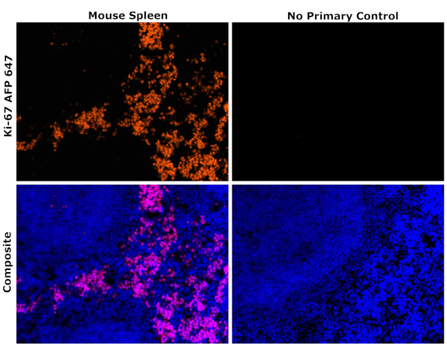
Immunohistochemical analysis of Ki-67 was performed using formalin-fixed paraffin-embedded mouse spleen tissue sections. To expose the target protein, heat-induced epitope retrieval was performed on de-paraffinized sections using eBioscience™ IHC Antigen Retrieval Solution - Low pH (10X) (Product # 00-4955-58) diluted to 1X solution in water in a decloaking chamber at 110 degree Celsius for 15 minutes. Following antigen retrieval, the sections were blocked with 2% normal goat serum in 1X PBS for 45 minutes at room temperature, followed by 10 minutes of blocking using Endogenous Biotin-Blocking Kit (Product # E21390) along with 1X PBS wash for 5 minutes. Sections were then probed with or without Ki-67 Recombinant Rat Monoclonal Antibody (SolA15), Alexa Fluor™ Plus 647 (Product # 740008TP647) at 1:100 dilution in 0.1% normal goat serum overnight at 4 degree Celsius in a humidified chamber. ReadyProbes™ Tissue Autofluorescence Quenching Kit (Product # R37630) was used to quench autofluorescence from the tissues. Nuclei were stained with DAPI (Product # D1306) and the sections were mounted using ProLong™ Glass Antifade Mountant (Product # P36984). The images were captured on EVOS™ M7000 Imaging System (Product # AMF7000) at 20X magnification.
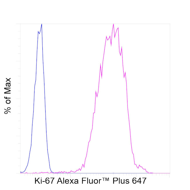
HeLa cells were fixed and permeabilized using the Foxp3 / Transcription Factor Staining Buffer Set (Product # 00-5523-00) and then stained intracellularly with 0.25 µg of Ki-67 Recombinant Rat Monoclonal Antibody (SolA15), Alexa Fluor™ Plus 647 (Product # 740008TP647) (pink histogram) or left unstained (blue histogram). Viable cells were used for analysis, as determined by Fixable Viability Dye eFluor™ 450 (Product # 65-0863-18). The flow cytometry data was acquired using Attune™ NxT Flow Cytometer (Product # A29004).
Antibody specificity was demonstrated by CRISPR-Cas9 mediated knockout of target protein. A loss of signal was observed for Ki-67 in KO cell line compared to control cell line using Ki-67 Recombinant Rat Monoclonal Antibody (SolA15), Alexa Fluor™ Plus 647 (740008TP647). {KO}