


 全部商品分类


RT KI67 PU RMAB

 下载产品说明书 下载COA 下载SDS
 用小程序,查商品更便捷


 收藏
收藏
 对比
对比 咨询
咨询种属反应
宿主/亚型
Expression System
分类
类型
克隆号
偶联物
形式
浓度
纯化类型
保存液
内含物
保存条件
运输条件
RRID
产品详细信息
Recombinant Rat monoclonal antibodies are produced using in vitro expression systems. Recombinant antibodies are produced using specific genes that code for the desired antibodies. These genes are cloned into an expression vector and expressed in vitro. The advantages of recombinant antibodies include better specificity and lot-to-lot consistency.
It is recommended that the antibody be carefully titrated for optimal performance in the assay of interest.
靶标信息
Ki-67 is a nuclear protein that is expressed during various stages in the cell cycle, particularly during late G1, S, G2, and M phases. The protein has a forkhead associated domain (FHA) through which it associates with euchromatin at the perichromosomal layer, the centromeric heterochromatin, and the nucleolus. Ki-67 is shown to have a cell cycle dependent topographical distribution with perinucleolar expression at G1, expression in the nuclear matrix at G2, and expression on the chromosomes during M phase. Ki-67 is commonly used as a proliferation marker because it is not detected in G0 cells, but increases steadily from G1 through mitosis. Ki-67 antibodies are useful in establishing the cell growing fraction in neoplasms. In neoplastic tissues, the prognostic value is comparable to the tritiated thymidine-labelling index. The correlation between low Ki-67 index and histologically low-grade tumors is strong. Ki-67 is routinely used as a neuronal marker of cell cycling and proliferation.
仅用于科研。不用于诊断过程。未经明确授权不得转售。
生物信息学
蛋白别名: Antigen identified by monoclonal antibody Ki-67; Antigen identified by monoclonal antibody Ki-67 homolog; Antigen KI-67; Antigen KI-67 homolog; Proliferation marker protein Ki-67; proliferation-related Ki-67 antigen; protein phosphatase 1, regulatory subunit 105; RP11-380J17.2
基因别名: D630048A14Rik; Ki-67; Ki67; KIA; MIB-; MIB-1; MKI67; PPP1R105
UniProt ID:(Human) P46013, (Mouse) E9PVX6
Entrez Gene ID:(Human) 4288, (Mouse) 17345
参考图片
Immunofluorescent analysis of Ki-67 was performed using 70% confluent HeLa cells. The cells were fixed with 4% paraformaldehyde for 10 minutes, permeabilized with 0.1% Triton X-100 for 15 minutes and blocked with 2% BSA for 1 hour at room temperature. The cells were stained with Ki-67 Recombinant Rat Monoclonal Antibody (SolA15) (Product # 740008T, 1:400) at 4 degree Celsius overnight and then labeled with Donkey Anti-Rat Secondary Antibody, Alexa Fluor™ 647 (Product # A78947) at a dilution of 1:2,000 for 1 hour at room temperature. Panel a) shows representative images of cells that were stained for detection and localization of Ki-67. Panel b) shows representative images of cells stained for nuclei using SYTOX™ Green Nucleic Acid Stain (Product # S7020, 1:10,000). Panel c) represents cytoskeletal F-actin staining using Alexa Fluor™ Plus 405 Phalloidin (Product # A30104, 1:300). The slides were mounted using ProLong™ Diamond Antifade Mountant (Product # P36961). Panel d) is a composite image of panels a, b, and c, clearly demonstrating nuclear localization of Ki-67. Panel e) represents no primary antibody control to assess the background. The images were captured at 60X magnification.
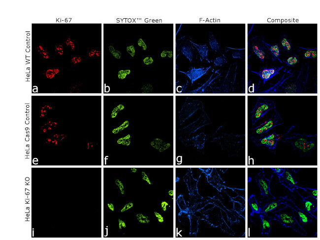
Knockout of Ki-67 was achieved by CRISPR-Cas9 genome editing using LentiArray™ Lentiviral sgRNA (Product # A32042, AssayID CRISPR955686_LV) and LentiArray Cas9 Lentivirus (Product # A32069). Immunofluorescence analysis was performed on wild type HeLa cells (panel a-d), HeLa Cas9 control cells (panel e-h) and HeLa Ki-67 KO cells (panel i-l). Cells were fixed, permeabilized, and labelled with Ki-67 Recombinant Rat Monoclonal Antibody (SolA15) (Product # 740008T, 1:400), at 4 degree Celsius overnight and then labeled with Donkey Anti-Rat Secondary Antibody, Alexa Fluor™ 647 (Product # A78947) at a dilution of 1:2,000 for 1 hour at room temperature. Nuclei (green) were stained using SYTOX™ Green Nucleic Acid Stain (Product # S7020, 1:10,000) and Alexa Fluor™ Plus 405 Phalloidin (Product # A30104, 1:300) was used for cytoskeletal F-actin (blue) staining. The slides were mounted using ProLong™ Diamond Antifade Mountant (Product # P36961). Loss of signal (panel i,l) upon CRISPR mediated knockout (KO) confirms that the antibody is specific to Ki-67 (red). The images were captured at 60X magnification.&(8)203;
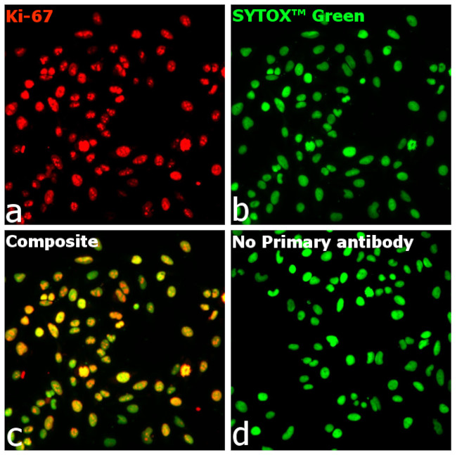
Immunofluorescent analysis of Ki-67 was performed using 70% confluent HeLa cells. The cells were fixed with 4% paraformaldehyde for 10 minutes, permeabilized with 0.1% Triton X-100 for 15 minutes and blocked with 2% BSA for 1 hour at room temperature. The cells were stained with Ki-67 Recombinant Rat Monoclonal Antibody (SolA15) (Product # 740008T, 1:1,000) at 4 degree Celsius overnight and then labeled with Donkey Anti-Rat Secondary Antibody, Alexa Fluor™ 647 (Product # A78947) at a dilution of 1:2,000 for 1 hour at room temperature. Panel a) shows representative images of cells that were stained for detection and localization of Ki-67. Panel b) shows representative images of cells stained for nuclei using SYTOX™ Green Nucleic Acid Stain (Product # S7020, 1:10,000). Panel c) is a composite image of panels a, and b, clearly demonstrating nuclear localization of Ki-67. Panel d) represents no primary antibody control to assess the background. The images were captured at 20X magnification using CellInsight™ CX7 LZR High-Content Screening (HCS) Platform.
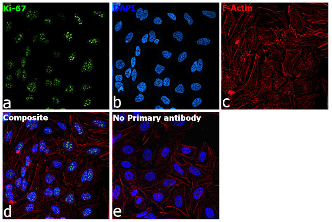
Immunofluorescence analysis of Ki-67 was performed using 70% confluent HeLa cells. The cells were fixed with 4% paraformaldehyde for 10 minutes, permeabilized with 0.1% Triton™ X-100 for 15 minutes and blocked with 2% BSA for 45 minutes at room temperature. The cells were labelled with Ki-67 Recombinant Rat Monoclonal Antibody (SolA15) (Product # 740008T, 1,10,000l) in 0.1% BSA, incubated at 4 degree Celsius overnight and then labelled with Donkey anti-Rat IgG (H+L) Highly Cross-Adsorbed Secondary Antibody, Alexa Fluor™ Plus 488 (Product # A48269, 1:2,000), for 45 minutes at room temperature (Panel a: Green). Nuclei (Panel b: Blue) were stained with ProLong™ Diamond Antifade Mountant with DAPI (Product # P36962). F-actin (Panel c: Red) was stained with Rhodamine Phalloidin (Product # R415, 1:300). Panel d represents HeLa cells showing nuclear localization. Panel e represents control cells with no primary antibody to assess background. The images were captured at 40X magnification.
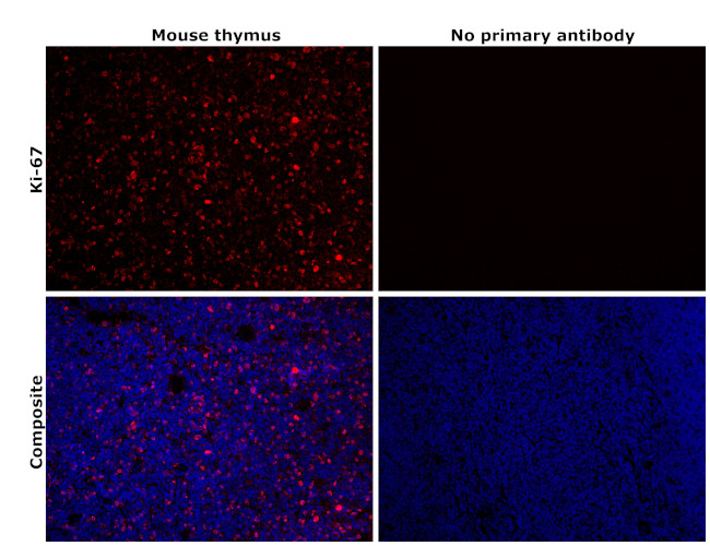
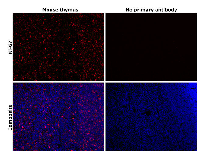
Immunohistochemical analysis of Ki-67 was performed using formalin-fixed paraffin-embedded mouse thymus tissue sections. To expose the target protein, heat-induced epitope retrieval was performed on de-paraffinized sections using eBioscience™ IHC Antigen Retrieval Solution - High pH (10X) (Product # 00-4956-58) diluted to 1X solution in water in a decloaking chamber at 110 degree Celsius for 15 minutes. Following antigen retrieval, the sections were blocked with 2% normal goat serum in 1X PBS for 45 minutes at room temperature and then probed with Ki-67 Recombinant Rat Monoclonal Antibody (SolA15) (Product # 740008T) at 0.5 µg/mL in 0.1% normal goat serum overnight at 4 degree Celsius in a humidified chamber. Detection was performed using Goat anti-Rat IgG (H+L) Highly Cross-Adsorbed Secondary Antibody, Alexa Fluor™ Plus 594 (Product # A48264, 1:2,000) in 0.1% normal goat serum for 45 minutes at room temperature. ReadyProbes™ Tissue Autofluorescence Quenching Kit (Product # R37630) was used to quench autofluorescence from the tissues. Nuclei were stained with DAPI (Product # D1306) and the sections were mounted using ProLong™ Glass Antifade Mountant (Product # P36984). The images were captured on EVOS™ M7000 Imaging System (Product # AMF7000) at 20X magnification and externally deconvoluted. The antibody stains specifically to the nucleus of cells in the thymus, as shown in the figure.
Immunohistochemical analysis of Ki-67 was performed using formalin-fixed paraffin-embedded mouse thymus tissue sections. To expose the target protein, heat-induced epitope retrieval was performed on de-paraffinized sections using eBioscience™ IHC Antigen Retrieval Solution - Low pH (10X) (Product # 00-4955-58) diluted to 1X solution in water in a decloaking chamber at 110 degree Celsius for 15 minutes. Following antigen retrieval, the sections were blocked with 2% normal goat serum in 1X PBS for 45 minutes at room temperature and then probed with Ki-67 Recombinant Rat Monoclonal Antibody (SolA15) (Product # 740008T) at 0.5 µg/mL in 0.1% normal goat serum overnight at 4 degree Celsius in a humidified chamber. Detection was performed using Goat anti-Rat IgG (H+L) Highly Cross-Adsorbed Secondary Antibody, Alexa Fluor™ Plus 594 (Product # A48264, 1:2,000) in 0.1% normal goat serum for 45 minutes at room temperature. ReadyProbes™ Tissue Autofluorescence Quenching Kit (Product # R37630) was used to quench autofluorescence from the tissues. Nuclei were stained with DAPI (Product # D1306) and the sections were mounted using ProLong™ Glass Antifade Mountant (Product # P36984). The images were captured on EVOS™ M7000 Imaging System (Product # AMF7000) at 20X magnification and externally deconvoluted. The antibody stains specifically to the nucleus of cells in the thymus, as shown in the figure.
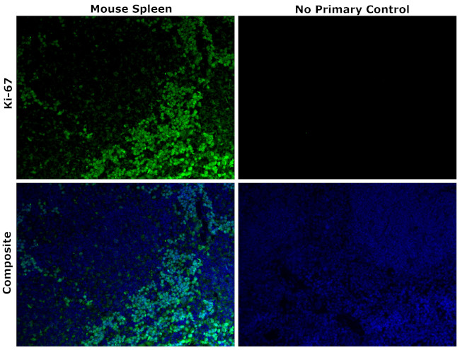
Immunohistochemical analysis of Ki-67 was performed using formalin-fixed paraffin-embedded mouse spleen tissue sections. To expose the target protein, heat-induced epitope retrieval was performed on de-paraffinized sections using eBioscience™ IHC Antigen Retrieval Solution - High pH (10X) (Product # 00-4956-58) diluted to 1X solution in water in a decloaking chamber at 110 degree Celsius for 15 minutes. Following antigen retrieval, the sections were blocked with 2% normal goat serum in 1X PBS for 45 minutes at room temperature and then probed with or without Ki-67 Recombinant Rat Monoclonal Antibody (Product # 740008T) at 1:200 dilution in 0.1% normal goat serum overnight at 4 degree Celsius in a humidified chamber. Detection was performed using Goat anti-Rat IgG (H+L) Cross-Adsorbed Secondary Antibody, Alexa Fluor™ 488 (Product # A11006) at a dilution of 1:2,000 in 0.1% normal goat serum for 1 hour at room temperature. ReadyProbes™ Tissue Autofluorescence Quenching Kit (Product # R37630) was used to quench autofluorescence from the tissues. Nuclei were stained with DAPI (Product # D1306) and the sections were mounted using ProLong™ Glass Antifade Mountant (Product # P36984). The images were captured on EVOS™ M7000 Imaging System (Product # AMF7000) at 20X magnification and externally deconvoluted.
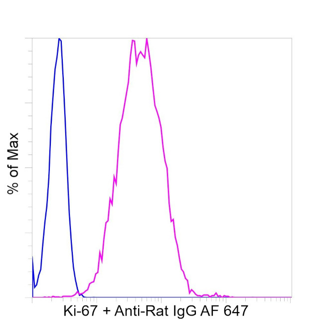
HeLa cells were fixed and permeabilized using the Foxp3 / Transcription Factor Staining Buffer Set (Product # 00-5523-00) and then stained intracellularly with 0.063 µg of Ki-67 Recombinant Rat Monoclonal Antibody (SolA15) (Product # 740008TM) (pink histogram) or 0.063 µg of Rat IgG2a kappa Isotype Control (Product # 14-4321-85) (blue histogram), followed by Donkey anti-Rat IgG (H+L) Highly Cross-Adsorbed Secondary Antibody, Alexa Fluor™ 647 (Product # A78947). Viable cells were used for analysis, as determined by Fixable Viability Dye eFluor™ 450 (Product # 65-0863-18). The flow cytometry data was acquired using Attune™ NxT Flow Cytometer (Product # A29004).
Antibody specificity was demonstrated by CRISPR-Cas9 mediated knockout of target protein. A loss of signal was observed for target protein in Ki-67 KO cell line compared to control cell line, using Ki-67 Recombinant Rat Monoclonal Antibody (SolA15) (Product # 740008T). {KO}