


 全部商品分类
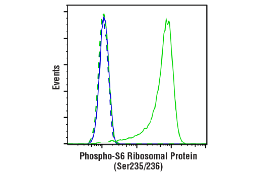


Phospho-S6 Ribosomal Protein (Ser235/236) (D57.2.2E) XP® Rabbit mAb (BSA and Azide Free)

 下载产品说明书 下载COA 下载SDS
 用小程序,查商品更便捷


 收藏
收藏
 对比
对比 咨询
咨询
Monoclonal antibody is produced by immunizing animals with a synthetic phosphopeptide corresponding to residues surrounding Ser235 and Ser236 of human ribosomal protein S6.
Product Usage Information
This product is the carrier free version of product #4858. All data were generated using the same antibody clone in the standard formulation which contains BSA and glycerol. This formulation is ideal for use with technologies requiring specialized or custom antibody labeling, including fluorophores, metals, lanthanides, and oligonucleotides. It is not recommended for ChIP, ChIP-seq, CUT&RUN, or CUT&Tag assays. If you require a carrier free formulation for chromatin profiling, please contact us. Optimal dilutions/concentrations should be determined by the end user.BSA and Azide Free antibodies are quality control tested by size exclusion chromatography (SEC) to determine antibody integrity.
Specificity/Sensitivity
Species Reactivity:
Human, Mouse, Rat, Monkey, Mink, S. cerevisiae
Store at -20°C. This product will freeze at -20°C so it is recommended to aliquot into single-use vials to avoid multiple freeze/thaw cycles. A slight precipitate may be present and can be dissolved by gently vortexing. This will not interfere with antibody performance.
参考图片
Flow cytometric analysis of Jurkat cells, untreated (green) or treated with LY294002 #9901, Wortmannin #9951, and U0126 #9903 (50 μM, 1 μM, and 10 μM, 2 hr; blue) using Phospho-S6 Ribosomal Protein (Ser235/236) (D57.2.2E) XP® Rabbit mAb (solid lines) or concentration-matched Rabbit (DA1E) mAb IgG XP® Isotype Control #3900 (dashed lines). Anti-rabbit IgG (H+L), F(ab')2 Fragment (Alexa Fluor® 488 Conjugate) #4412 was used as a secondary antibody. Data were generated using the standard formulation of this product.
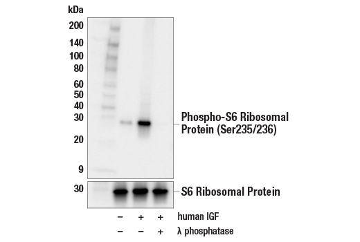
Western blot analysis of extracts from serum-starved MCF7 cells, untreated (-) or treated (+) with combinations of the following treatments as indicated: human IGF-1 (100 ng/mL, 10 min) and λ phosphatase, using Phospho-S6 Ribosomal Protein (Ser235/236) (D57.2.2E) XP® Rabbit mAb (upper) or S6 Ribosomal Protein (5G10) Rabbit mAb #2217 (lower).
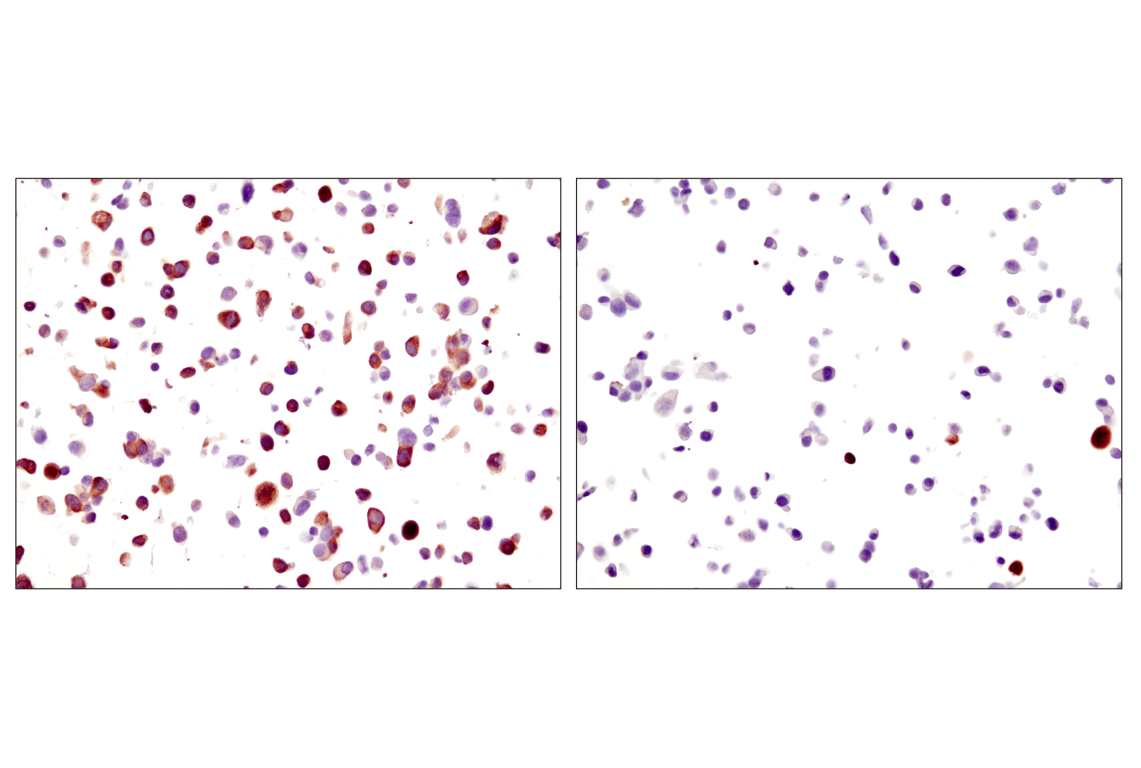
Immunohistochemical analysis of paraffin-embedded LNCaP cells, untreated (left) or LY294002-treated (right), using Phospho-S6 Ribosomal Protein (S235/236) (D57.2.2E) XP® Rabbit mAb on SignalSlide® Phospho-Akt (Ser473) IHC Controls #8101. Data were generated using the standard formulation of this product.
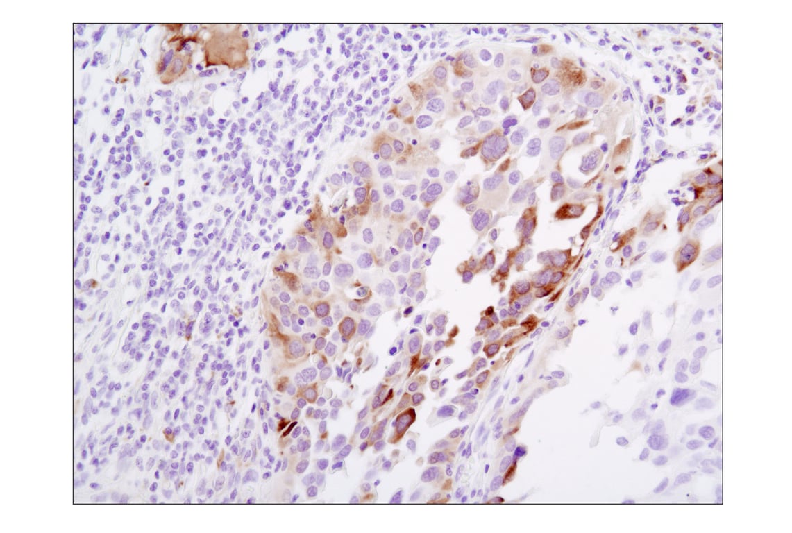
Immunohistochemical analysis of paraffin-embedded human lung carcinoma using Phospho-S6 Ribosomal Protein (Ser235/236) (D57.2.2E) XP® Rabbit mAb. Data were generated using the standard formulation of this product.
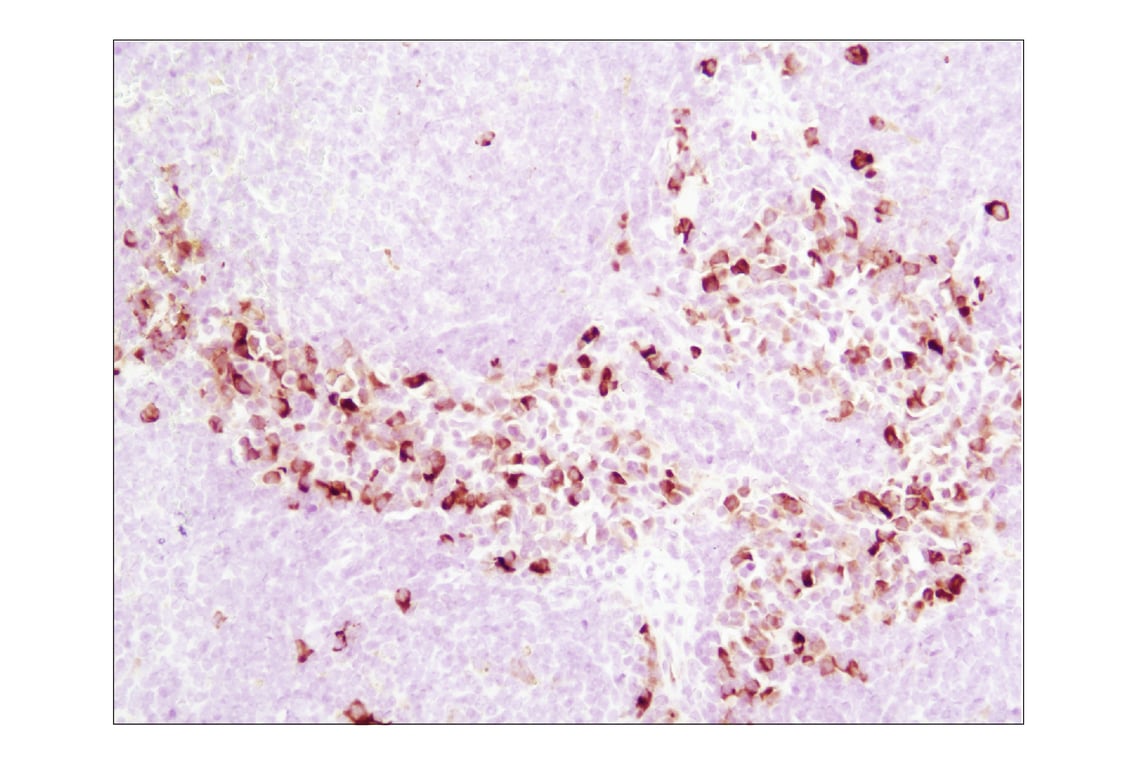
Immunohistochemical analysis of paraffin-embedded mouse spleen using Phospho-S6 Ribosomal Protein (Ser235/236) (D57.2.2E) XP® Rabbit mAb. Data were generated using the standard formulation of this product.
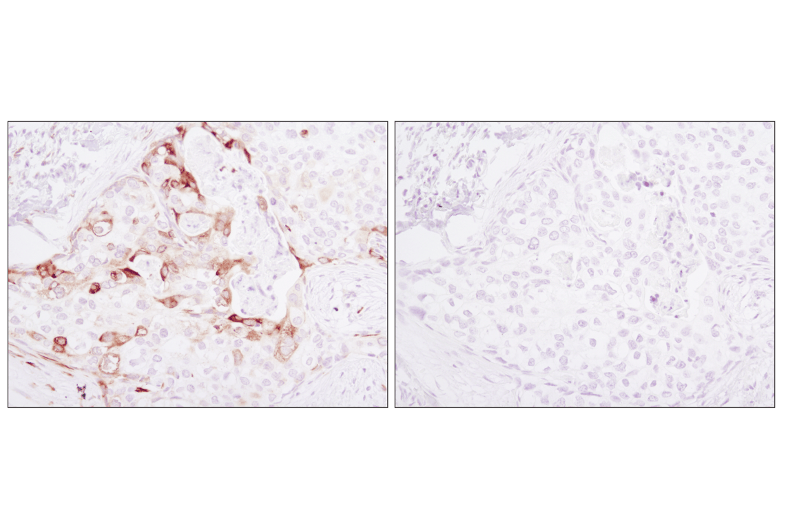
Immunohistochemical analysis of paraffin-embedded human breast carcinoma using Phospho-S6 Ribosomal Protein (Ser235/236) (D57.2.2E) XP® Rabbit mAb in the presence of control peptide (left) or Phospho-S6 Ribosomal Protein (Ser235/236) Blocking Peptide #1220 (right). Data were generated using the standard formulation of this product.
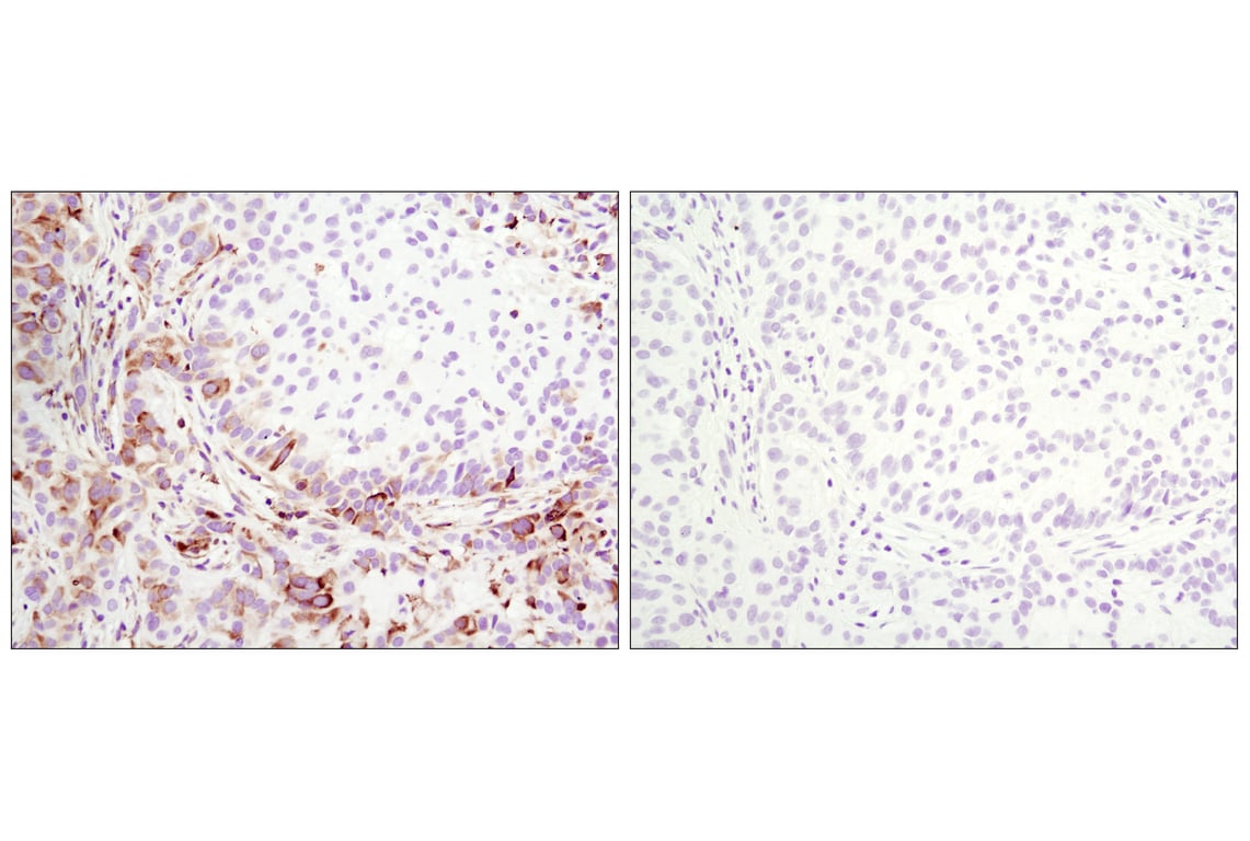
Immunohistochemical analysis of paraffin-embedded A549 xenograft, untreated (left) or λ phosphatase-treated (right), using Phospho-S6 Ribosomal Protein (Ser235/236) (D57.2.2E) XP® Rabbit mAb. Data were generated using the standard formulation of this product.
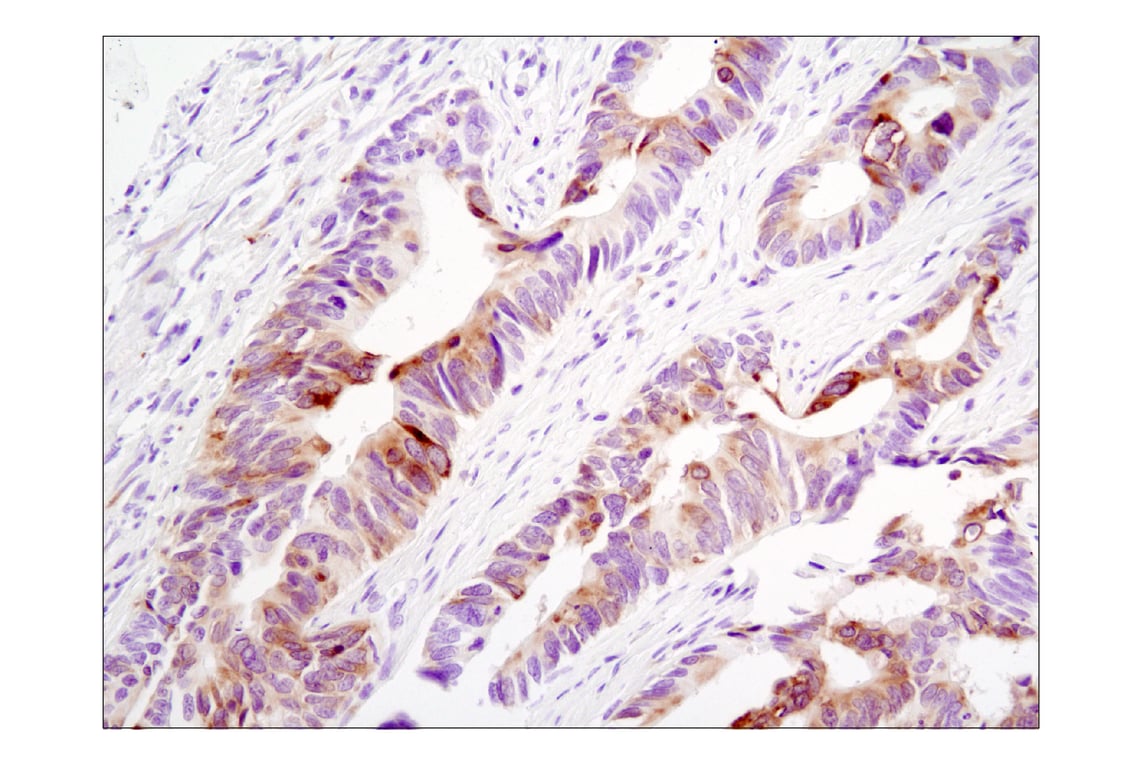
Immunohistochemical analysis of paraffin-embedded human colon carcinoma using Phospho-S6 Ribosomal Protein (Ser235/236) (D57.2.2E) XP® Rabbit mAb. Data were generated using the standard formulation of this product.
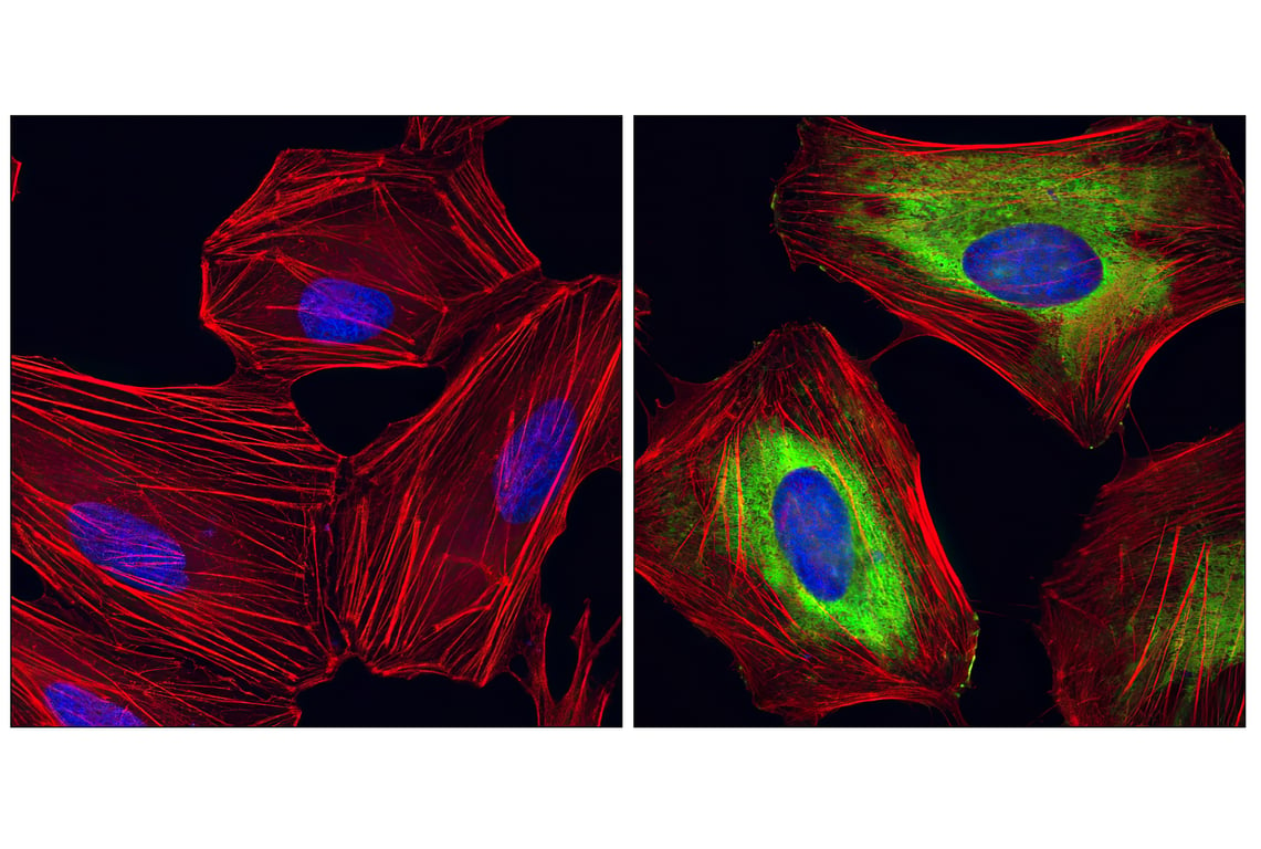
Confocal immunofluorescent analysis of HeLa cells, rapamycin-treated (left) or 20% serum-treated (right), using Phospho-S6 Ribosomal protein (Ser235/Ser236) (D57.2.2E) XP® Rabbit mAb (green). Actin filaments have been labeled with Alexa Fluor® 555 phalloidin (red). Blue pseudocolor = DRAQ5® #4084 (fluorescent DNA dye). Data were generated using the standard formulation of this product.